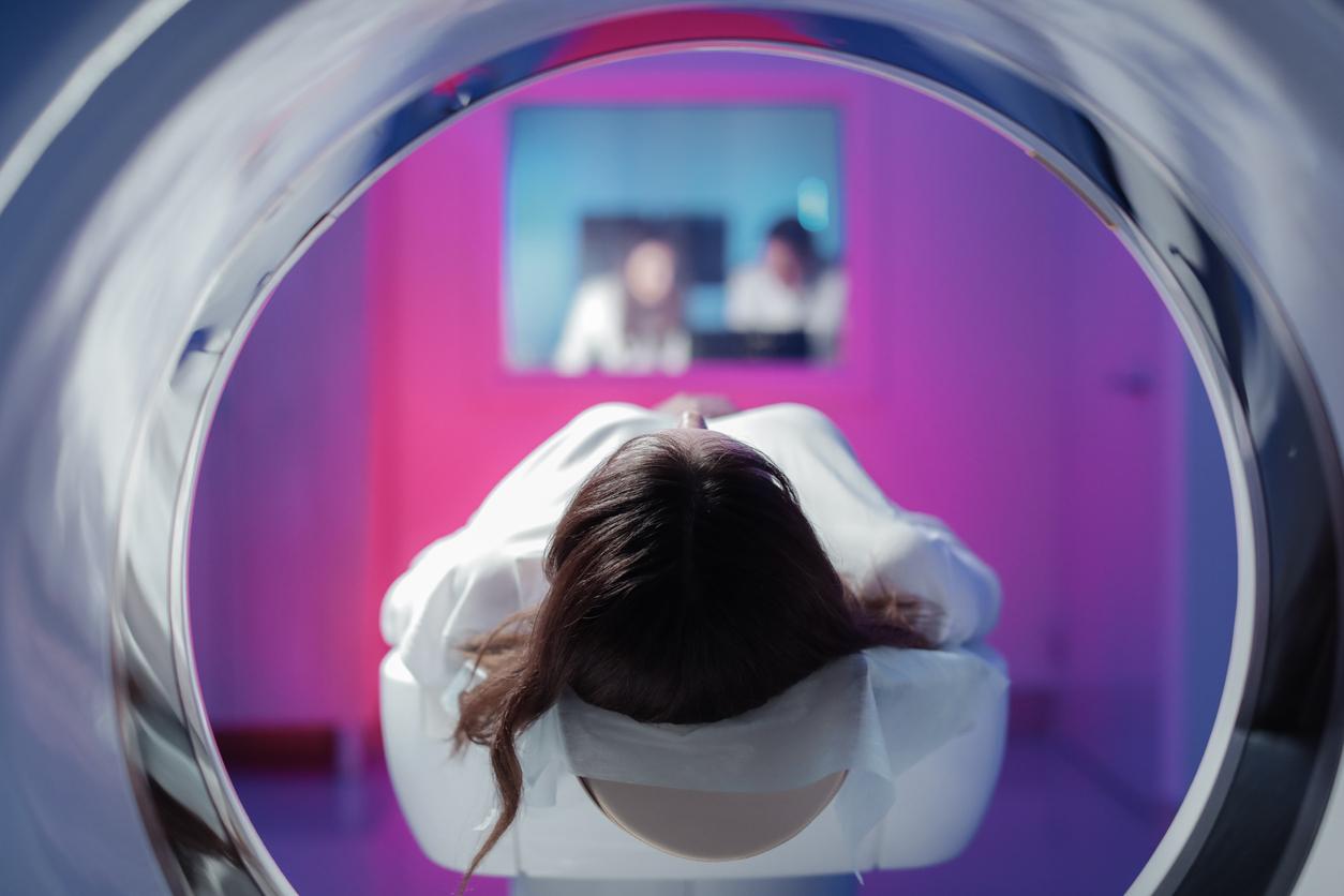
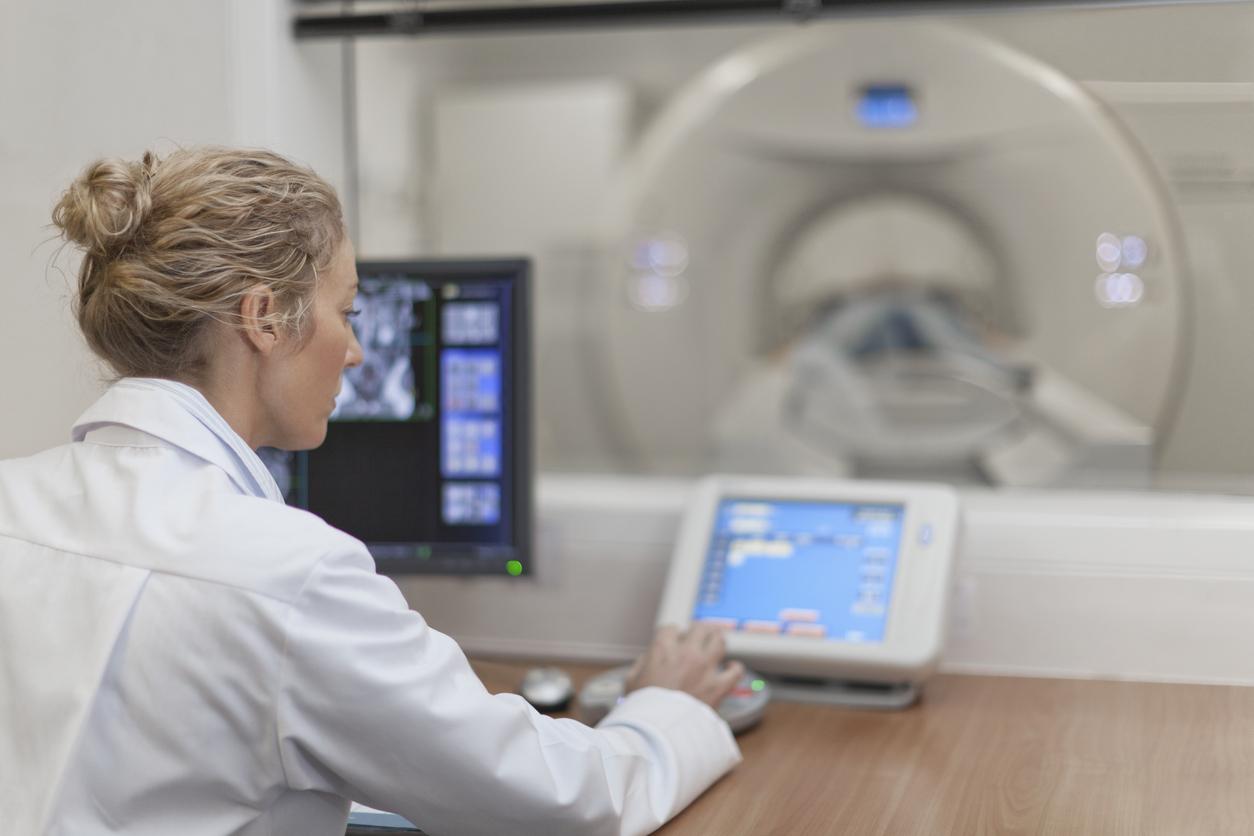
At the last French radiology congress, the 3-dimensional scanner was presented. We have clearly passed the stage of a simple black and white photograph. Frankly, it’s very impressive, the images come out of the screen and are suddenly visible to several people at the same time, and without glasses. This technology is applicable to both CT and MRI examinations. And both an ankle and a cerebral aneurysm can burst the screen.
The question that immediately comes to mind is: gadget or not?
In any case, thanks to this system, the organs are visible from eight viewing angles, 10 meters away, and with a very wide angle of diffusion. Concretely, this makes it possible, for example, to visualize the rib cage with all the organs and vessels. Afterwards, the fact that several people can see these images at the same time is certainly a bit like home cinema… But, in fact, it allows the entire medical team to prepare for the operation.
In addition, these relief images are also of interest to the patient: they are much more meaningful than the cross-sectional images of scanners. It is often said that a drawing is better than a long speech… Obviously, a drawing that has a significant price. All the equipment would cost around 12,000 euros. So, in times of budgetary restriction, it will probably be necessary to wait before medical imaging switches to the 3rd dimension. Or maybe it should be reserved for very specific situations where 3D is a real plus.
A scenario similar to what happened with the arrival of 3D ultrasounds. Initially, parents who could afford it all wanted to see their babies in 3D. Doctors put it all down, seeing it as a luxury gadget. And today, this technique is mainly used in case of suspicion of physical anomaly, in particular to diagnose a harelip.
And then, while technology is advancing, there is still a dire shortage of sufficient imaging devices. You still have to wait 32 days to have access to an MRI. We are therefore far from the goal of the cancer plan, which was ten days …
.


