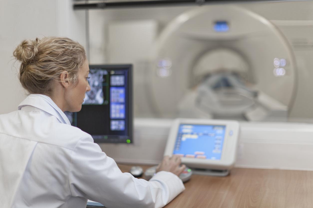
A new MRI method makes it possible to see how air circulates in the lungs, and therefore makes it possible to assess the effects of asthma treatments in real time.

- A new MRI method can see how air enters and leaves the lungs precisely and in real time.
- This makes it easier to assess the effects of treatment on patients’ lung function.
- This new method can also be useful for patients who have had a lung transplant. It makes it possible to identify early signs of rejection.
Researchers from the University of Newcastle have made a discovery which will greatly improve the care of people with respiratory disorders such as asthma or chronic obstructive pulmonary disease (COPD) as well as patients who have had a lung transplant.
They developed a new MRI method that can see how air enters and leaves the lungs when people breathe, and therefore accurately assess their lung function in real time. Their discovery was presented in two scientific journals.
Asthma: a gas makes it possible to check the effectiveness of the treatment in real time
In an article published in the journal Radiology, British scientists reveal that they have demonstrated that a gas called perfluoropropane is visible during MRI scans of the lungs, and improves the quality of the exams. Indeed, this substance, which can be breathed safely by patients, makes it possible to accurately see the circulation of air in the pulmonary system.
“Our scans show areas of uneven ventilation in patients with lung disease and tell us which parts of the lungs are improving with treatment. For example, when we examine a patient while they are taking their lung disease medications. asthma, we can see which areas of his lungs are best at moving air in and out with each breath.”explains Professor Pete Thelwall who led the research in a press release.
For researchers, knowing precisely which part of the organ is well ventilated and which is not will improve the care of asthmatic patients and the evaluation of the effectiveness of their treatments. They add that this new imaging method will also “valuable in clinical trials of new drugs for lung disease”.
Lung: an MRI method also useful for transplant patients
This new MRI method is not only useful for monitoring patients with asthma or COPD. A second study published in JHLT Openshows that it can also be interesting for patients who have had a lung transplant.
Researchers say the sensitivity of the measurement offered by this technique can help doctors spot early changes in lung function in transplant patients. This allows them to identify respiratory problems or signs of rejection early.
“We hope that this new type of scanner will allow us to see changes in transplanted lungs earlier and before signs of damage are present in usual blowing tests. This would allow any treatment to begin earlier and help protect transplanted lungs against further damage”explains the co-author of the study, Professor Andrew Fisher.


