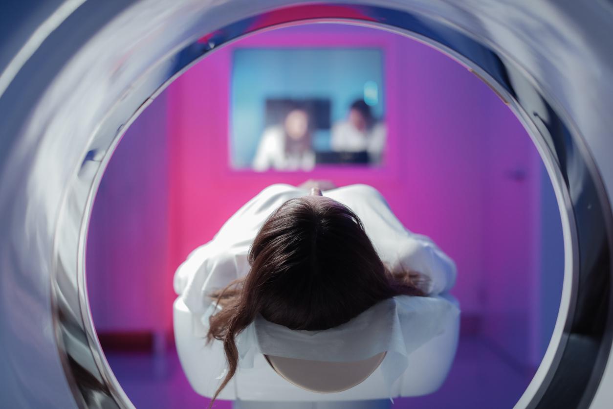
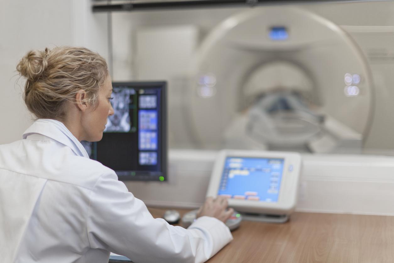
Using magnetic resonance imaging (MRI), researchers have realized that it is possible to predict post-traumatic stress disorder by detecting its biomarkers after traumatic brain injury.
- Still today difficult to prevent, post-traumatic stress disorder would affect the volume of several regions of the brain.
- This new study shows that it is possible to detect these biomarkers after traumatic brain injury using a brain scan.
Affecting about one in ten French people, post-traumatic stress disorder (PTSD) refers to a set of reactions occurring in people who have experienced a traumatic event. Victims of attacks, torture, rape, serious accidents, aggression and veterans are thus particularly affected by this condition, which encompasses various symptoms. Most people who have developed PTSD suffer from sleep disorders (insomnia, nightmares), depression, develop addictions, a feeling of irritability, intense fear, anger, which can sometimes lead to outbursts of violence.
Although better understood and supported today, post-traumatic stress disorder remains a complex psychiatric disorder that is sometimes difficult to prevent or detect. A new study published in the journal Biological Psychiatry: Cognitive Neuroscience and Neuroimaging could, however, help doctors better predict the development of PTSD. According to its authors, the use of magnetic resonance imaging (MRI) could uncover potential brain biomarkers of PTSD in people with traumatic brain injury.
A lower volume of certain regions of the brain
To investigate the link between post-traumatic stress disorder and traumatic brain injury, the researchers used data from TRACK-TBI, a large longitudinal study of patients who present to the emergency room with traumatic brain injury severe enough to warrant CT. -scan (computed tomography).
“MRI studies conducted within two weeks of injury were used to measure the volumes of key brain structures thought to be involved in PTSD, explains Doctor Murray Stein, from the University of California at San Diego (United States). We found that the volume of several of these structures was predictive of PTSD three months after injury.”
In total, more than 400 patients with traumatic brain injury were followed and had a CT-Scan after their injury, then within 3 and 6 months to assess the risk of PTSD. After 3 months, 77 participants (18%) probably suffered from PTSD. After 6 months, 70 participants (16%) suffered from it.
Researchers found lower volume in brain regions called the cingulate cortex, superior frontal cortex, and insula, which predicted PTSD at 3 months. These regions are associated with arousal, attention and emotional regulation. Imaging, on the other hand, did not predict PTSD at 6 months.
A brain reserve against PTSD
Previous work had already shown lower volume in several of these brain regions in people with PTSD. For the authors of the study, these new results suggest that a “brain reserve”or higher cortical volumes, may provide some resistance against PTSD.
Although the biomarker for brain volume differences is not yet sufficiently substantiated to provide clinical indications, Dr. Stein believes that “This paves the way for future studies to look more closely at how these brain regions may contribute to or protect against mental health issues such as PTSD”.

.


