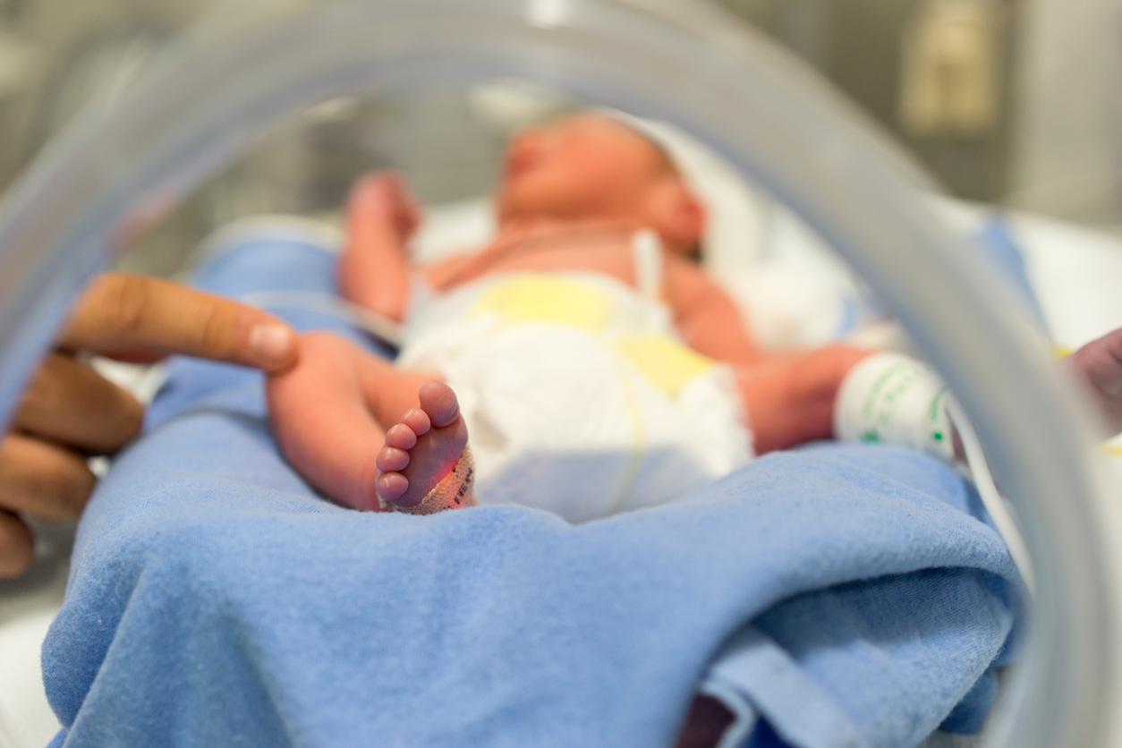
An international group of researchers determined when during pregnancy the pelvis takes shape and identified the genes responsible for this process.
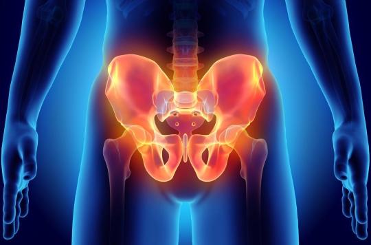
- The pelvis is made up of three bones: the ischium, ilium and pubis.
- Thanks to the pelvis, human beings do not have to move their body mass forward and use their joints to walk or balance.
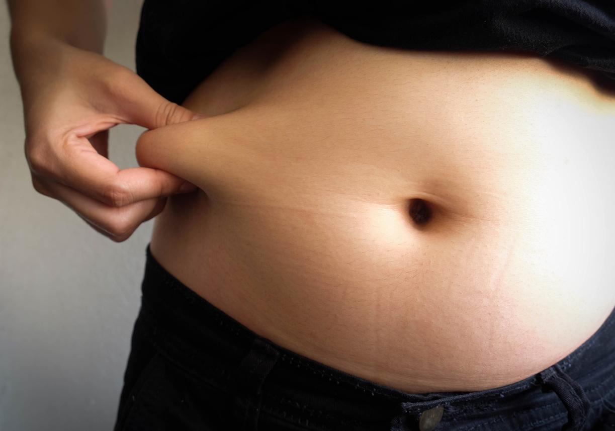
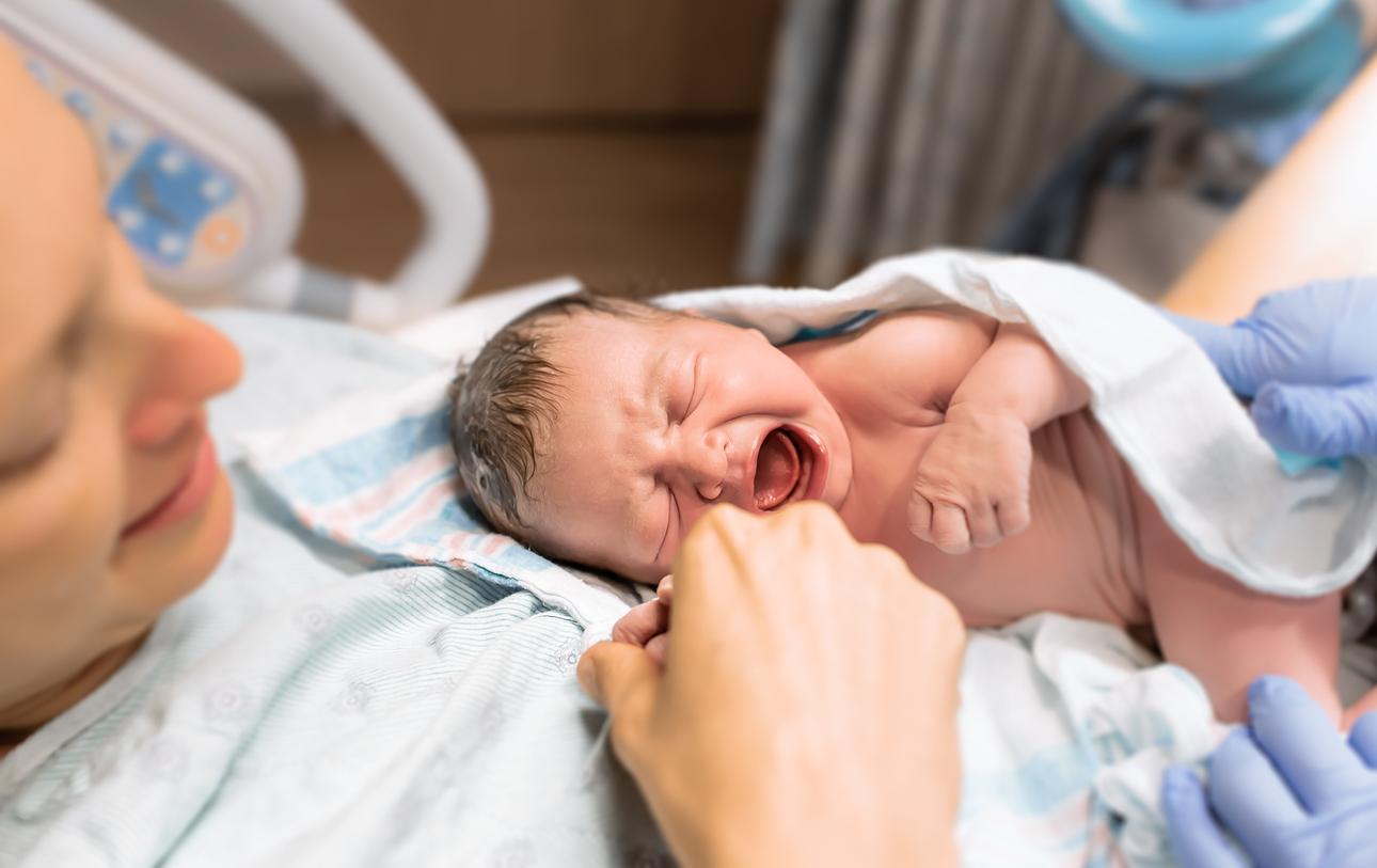
The pelvis is the lower part of the belly. This bony belt serves to join the lower limbs, the spine and the hip joint. Also, it contains the bladder, rectum and reproductive organs. The pelvis allows human beings to walk upright and women to give birth to children.
Pregnancy: the pelvis forms around the 6th or 8th week
Anatomically, the pelvis is well known. However, knowledge is limited when it comes to how and when this important bone structure takes shape during fetal development, according to an international team of scientists. This is why they decided to carry out a study published in the journal Science Advances.
As part of the work, the authors examined embryos aged 4 to 12 weeks under a microscope, with the consent of people who had legally terminated their pregnancies. They then compared samples of the developing human pelvis with mouse models to identify genes that trigger pelvic formation. They discovered hundreds of genes that are turned on or off around the sixth or eighth week of pregnancy.
Pelvis: a blockage allows the cartilage to continue to develop
According to the findings, the formation of the pelvis occurs while the bones are still cartilage, allowing them to bend, rotate, expand and grow easily. Unlike other cartilage in the body, this developing pelvic structure stays in the form of cartilage for a longer time. “It appears that a blockage is occurring that allows the cartilage to continue to grow,” said Terence D. Capellini, author of the research, in a statement.


