Your body turned inside out
An indefinable pain or vague complaints. It is not always possible to see what is wrong with it from the outside of our body. Yet we want to know where the pain comes from or what causes the complaints. Fortunately, technology helps us turn the body inside out.
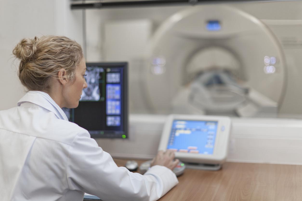
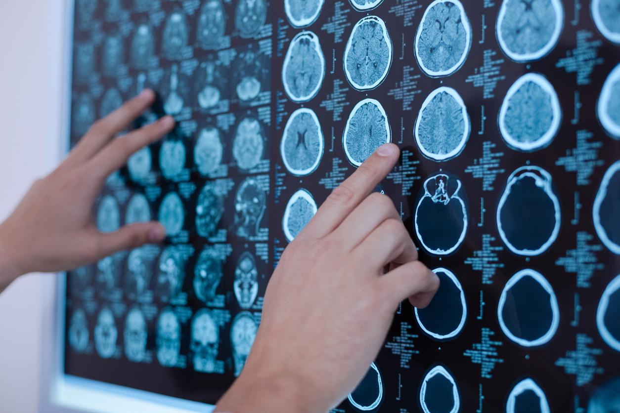
Imaging studies show what the body looks like from the inside. It is an important diagnostic tool. It also shows how extensive any deviations are. There are different types of scans that use different techniques. Each scan has its own specialty, but also its limitations.
- MRI scan
- CT-scan
- Ultrasound/sonography
- X-ray
- DXA scan
-
Nuclear medicine examination (PET scan, lung scan, bone scan)
MRI scan
An MRI scan provides an accurate picture of bones, joints and surrounding tissue such as muscles, tendons and cartilage. MRI stands for Magnetic Resonance Imaging. It is a scanning technique that uses magnetic waves and radio waves.
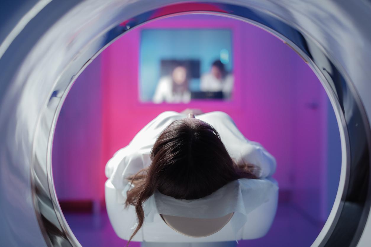
During the examination you will be in a narrow tunnel. The front and back are open. The device makes a thumping sound, so you will be given headphones and you can bring your own CD. It is important to stay still. Depending on the area to be surveyed, the survey will take between 30 and 90 minutes.
You will not feel anything from the magnetic waves. However, lying still for a long time can be uncomfortable and some people experience fear in the narrow tunnel. If you are pregnant less than 12 weeks or if you are suspected of being pregnant, it is advisable to consult your doctor first.
CT-scan
A CT scan (Computer Tomography) is an imaging study in which cross-sections of the body are made with X-rays. The computer reassembles the pictures into a whole.
Each shot is a cross-section of the body. The movable table on which you lie moves a few millimeters further under the arc where the scanner is located. This continues until the entire body part to be examined has been imaged.
In order to better visualize certain abnormalities, a contrast agent is sometimes used. This enters the body through the bloodstream. Most people hardly notice it, at most a warm feeling, but sometimes someone is hypersensitive to these substances.
By making the CT scan you feel nothing. It is also the intention here that you lie still. The position of your body and the duration of the scan depend on which part of the body needs to be imaged.
Ultrasound/sonography
Ultrasound uses sound waves to image tissues and organs. In this way we can gain insight into size, structure and any pathological abnormalities. The ultrasound is also used, for example, in prenatal examinations.
During the examination, a jelly is applied to bare skin. The researcher moves a small device over the skin that emits sound waves that are inaudible to us. These waves are reflected by tissues and converted into images that can be viewed directly on a screen.
Ultrasound is painless, only the conductive gel may feel a bit cold.
X-ray
In X-rays, bones and cartilage are made visible using X-rays. X-rays have a short wavelength and high penetrating power. They mainly make visible tissues that contain calcium.
Organs and blood vessels can also be visualized using contrast dye. Often the liquid contains iodine, because iodine can absorb X-rays.
Usually several photos are taken, in different directions. X-rays are reviewed by a radiologist.
The examination is not burdensome: you do not feel the radiation. The dose of X-rays required for the examination is small. An adult person is not harmed by this. However, radiation exposure to unborn children is not without danger; In case of pregnancy, therefore, always consult with the doctor.
DXA scan
With Dual Energy X-Ray Absorptiometry (DXA or DEXA) the calcium content in the bones is measured. In this way it can be investigated whether there is osteoporosis, or bone loss.
The DXA scan measures bone density using X-rays. The amount of calcium indicates whether you have osteoporosis and how likely it is that you will develop osteoporosis. This scan is also not burdensome.
Nuclear Medicine Research
In nuclear medicine research, abnormalities in tissue are visualized using a small amount of radioactive material and a special gamma camera. The places where the radioactive substance has accumulated are then made visible.
For example, a tumor has a faster metabolism and the diseased tissue will therefore absorb more dust and emit radiation. The gamma camera records this radiation.
The radioactive substance is usually injected into the bloodstream, but sometimes the substance is given orally, instilled, inhaled, or eaten. This depends on the body part or body function to be imaged. After insertion, the radioactive substance needs time to act. This can take anywhere from a few minutes to a few hours.
The amount of radiation released during a nuclear medicine examination is comparable to the dose you receive when taking X-rays. The radioactive substances leave the body again through the urine. Drinking a lot will speed up this process.
However, it is not recommended to take small children on your lap on the day of the examination. When breastfeeding, the milk should be expressed and washed out for the first 24 hours. The radiation can be harmful to young children.
The most common types of nuclear medicine examination are bone scan, PET scan, and lung scan.
Sources):


