Researchers have developed an ultrasound imaging method which aims to replace endoscopy.

A recent discovery could sound the death knell for endoscopy. Maysam Chamanzar and Matteo Giuseppe Scopelliti, respectively assistant professor and doctoral student at Carnegie Mellon University in Pittsburgh in the United States, have developed an ultrasound imaging method which aims to replace endoscopy. Their contributions were published in the journal Light: Science & Applications.
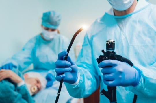
Endoscopy is the method most frequently used in medical imaging, in particular for the diagnosis of diseases of the lungs, colon, throat or intestines. During this procedure, doctors insert an endoscope, a long tube with a camera and light attached, through a small opening (the mouth or an incision made by a surgeon).
Endoscopy is an invasive and painful method that can leave sequelae such as significant sedation, cramps, chronic pain and sometimes tissue perforation and internal bleeding.
Replace the physical optical lens with a virtual lens
The two researchers explain in their paper that the tissues of the human body, being thick and opaque, limit the possibilities in terms of medical imaging. Tissues are made of large particles and membranes, which restrict the range and resolution of optical imaging. “Particularly in the visible infrared region of the spectrum,” explain the two researchers.
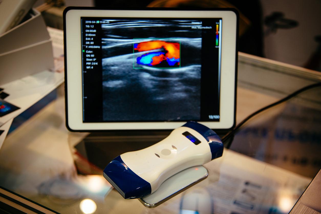
This new technique, on the contrary, uses ultrasound to design a “virtual lens” in the body rather than inserting a physical lens. The doctor can then adjust this lens by “changing the ultrasound pressure inside the medium” and take deep pictures that were not previously accessible by non-invasive techniques.
“When the ultrasound waves propagate in the medium, they alter its density and therefore its local index of refraction, explain the researchers. The medium is compressed in regions of high pressure and therefore high density, while it is rarefied in regions of negative pressure where the local density is reduced.
Additionally, adjusting and reconfiguring the ultrasound from the outside allows the lens to be moved within the medium through multiple tissue regions and thus image at different depth levels.
A method that could “revolutionize biomedical imaging”
This method could be used in brain imaging, in the diagnosis of skin diseases and in the identification of tumors in different organs and could be accompanied by a tool to be held in the hand or a patch on the skin. .
By simply applying it to the skin, healthcare professionals could obtain internal images without the potential side effects and pain of endoscopy.
“Being able to relay images of organs like the brain without the need to insert optical components provides an important alternative to implanting endoscopes in the body,” explains Maysam Chamanzar. This method can revolutionize the field of biomedical imaging.”

.


