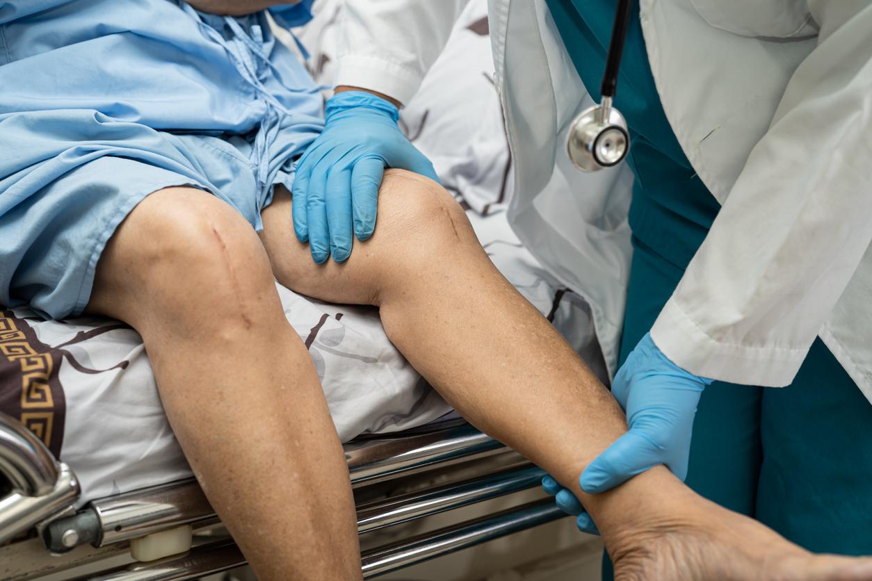
A person’s risk of suffering a meniscus tear could be revealed through radiomics, which is the assessment of various characteristics from imaging tests.
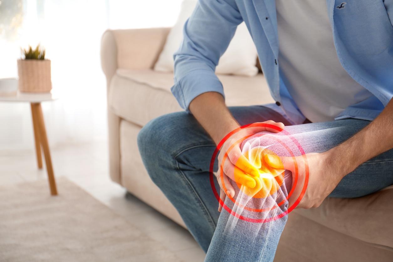
- Radiomics is a discipline that involves computer analysis of medical images and translating them into complex quantitative data.
- In a recent study, it made it possible to identify and classify 24 of 34 cases of meniscus tears.
- This technique identified a large area with high gray level, i.e. coarser texture, of the posterior horn as the most important variable.
Radiomics is a discipline, the concept of which has existed for around 40 years, but which has recently experienced an explosion. She “consists of the extraction of a large number of characteristics or measurements from medical images and, further, their exploitation to construct classification or prediction models”has explained Irene Buvatphysicist and director of the Laboratory for Translational Imaging in Oncology at the Institut Curie.
The meniscus of 215 adults was intact at the start of the study
A recent study focused on using radiomics, which reveals imperceptible patterns in medical images, to classify people who will develop a torn knee meniscus. To carry out the work, researchers from Michigan State University (United States) used magnetic resonance images (MRI) of 215 people whose menisci were intact at the time of registration. They assessed the condition of the crescent-shaped cartilages four years later. “A reader manually segmented the anterior and posterior horn of the medial meniscus of each participant during the medical visit. Then, 61 different radiomic features were extracted from each horn of the medial meniscus,” the team said.

Knee meniscus tear: radiomics identified 24 of 34 cases
According to the results, published in the journal Journal of Orthopedic Research, 34 participants developed meniscus tears. The use of radiomics at the start of the work made it possible to correctly identify and classify 24 of these 34 cases and 172 of the 181 controls with a sensitivity of 70.6% and a specificity of 95.0%. The authors explained that the classification and regression tree identified a large area with high gray level, i.e. coarser texture, of the posterior horn as the most important variable for classifying people who are at risk of developing a meniscus tear.
“This technique therefore provides sensitive and quantitative measurements of meniscus alterations that could help clinicians know when to intervene to guard against meniscus tears,” concluded the scientists.


