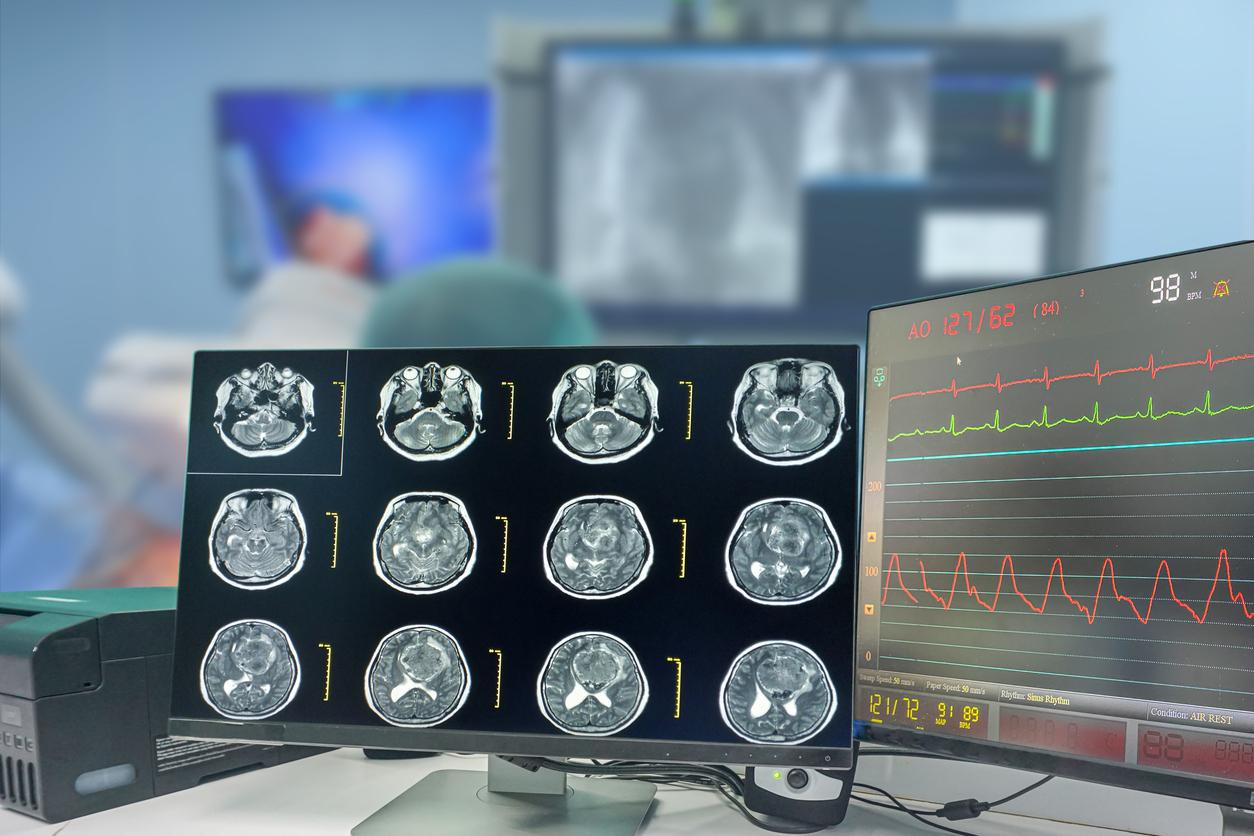
Researchers have developed a new method for analyzing 3D images in the context of glioblastoma biopsy. This breakthrough could lead to improved diagnosis and treatment.
-1613836465.jpg)
- Researchers have perfected a 3D biopsy technique which makes it possible to assess the interior of the tumor and the location of the different types of cells.
- This technique makes it possible to refine the diagnosis and thus to personalize the treatments to target the malignant cells.
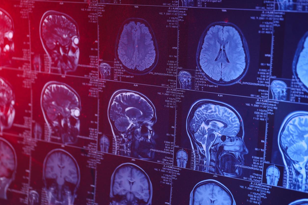
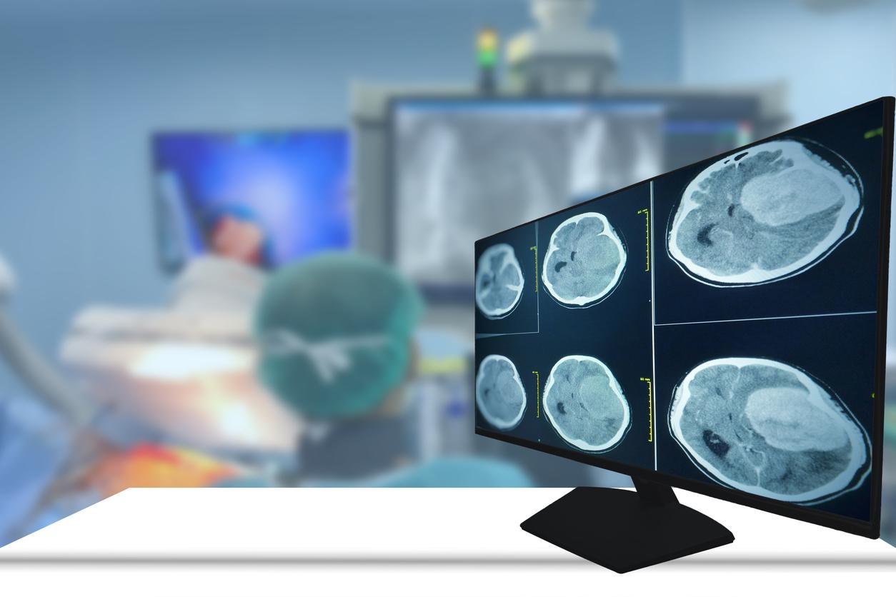
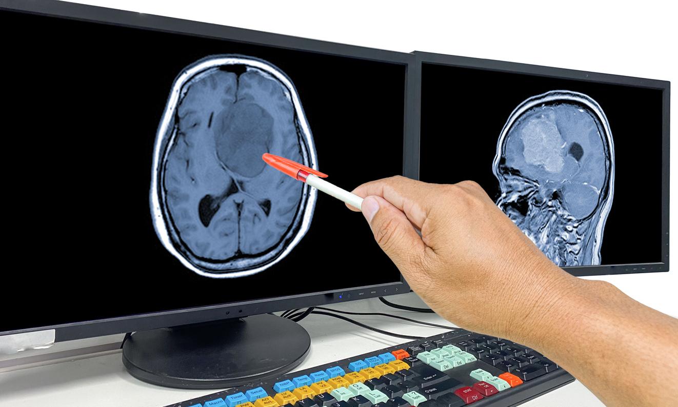
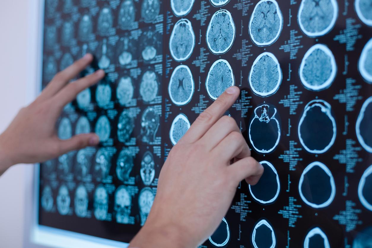
Glioblastoma is a malignant brain tumor that usually affects adults between the ages of 45 and 70. To diagnose it, doctors first prescribe a complete neurological examination to determine the level of functioning of the nervous system, where the tumor is located. Depending on the result of this examination, a scanner and Magnetic Resonance Imaging (MRI) of the brain may be requested to confirm the diagnosis and help determine the precise area where the tumor is located. Once located, the biopsy – the removal of one end of the glioblastoma – confirms the diagnosis. Researchers from the Institute of Neurosciences of the Autonomous University of Barcelona have just developed a new method of 3D biopsy of glioblastoma, which could make it possible to refine the diagnosis and, in the long term, to open up new therapeutic avenues.
3D biopsy to better appreciate the inside of the tumor
The work of the scientists was published in the journal Acta Neuropathologica Communications. With this new method of analyzing 3D images and quantitative data, the authors believe that it will be possible to better appreciate the interior of the tumor and the location of the different cell types. “It provides more comprehensive information than the 2D scans typically performed for neuropathological diagnosis.” assures George Paul Cribaro, first author of the study. With this technique, it would be possible to see several crucial elements to refine the diagnosis and better target treatments. Among them, the alterations of the tumoral blood vessels and the potential abnormalities of the vascular wall, which can hinder the entry of T-lymphocytes. They are the ones who can defend the body against tumor cells. Determining these vascular wall defects would make it possible to better personalize treatments and target malignant cells. This discovery is therefore an aid to the diagnosis but also, in the long term, to the treatments that will be put in place.
Differentiate the different areas of the tumor
Thanks to these 3D images, the authors also believe that they could differentiate between the two areas of the tumor. The tumor tissue on the one hand, and the stroma on the other hand, which supports the tumor and in which there are different immunological microenvironments. “This differentiation (…) could help in the diagnosis and in the search for new therapeutic targets”, says Carlos Barcia, works coordinator. These 3D biopsies will make it easier to understand the complexity of glioblastomas. Armed with this information, doctors will eventually be able to design new therapeutic approaches.
.


