An international study published in Nature Medicine shows that the tau protein, responsible for Alzheimer’s disease, spreads in four distinct patterns that lead to different symptoms with different prognoses in those affected.

- By combining the use of an algorithm and that of PET imaging, the researchers succeeded in identifying four subtypes of Alzheimer’s disease.
- These four subtypes are characterized by accumulation of tau protein in different regions of the brain, resulting in different symptoms and prognoses.
- This discovery could lead to the implementation of individualized treatments for patients affected by Alzheimer’s.
Affecting 900,000 people in France, Alzheimer’s disease is the most common form of age-related dementia. Still incurable at present, this neurodegenerative disease slowly, progressively and irreversibly leads to the dysfunction and then the death of nerve cells in the brain.
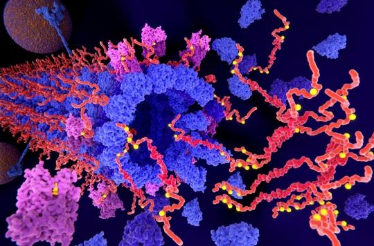
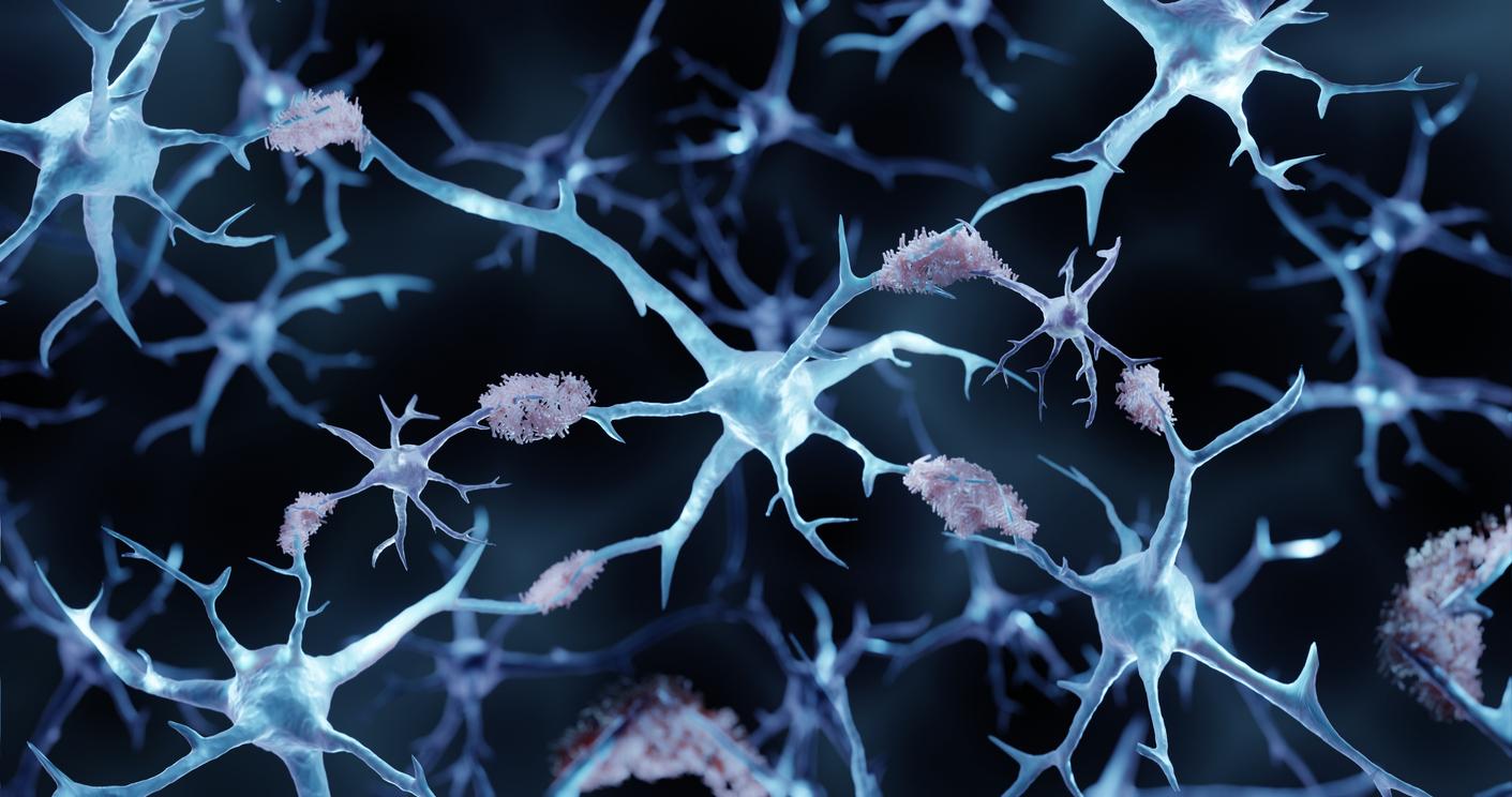
Two phenomena are involved. The disease begins with the formation of amyloid plaques in the brain, which accumulate abnormally in the neurons, leading to progressive and irreversible brain damage. This is followed by an abnormal accumulation of tau protein, which obstructs the interior of neurons.
In a study published in the journal NatureMedicinean international team of researchers show that this tau protein spreads in four distinct patterns. “This suggests that Alzheimer’s disease is an even more heterogeneous disease than previously thought, explains Jacob Vo-gel of McGill University, lead author of the study. We now have reason to re-evaluate the concept of typical Alzheimer’s and, in the long term, the methods we use to assess disease progression.”
4 diagrams with different symptoms
The spread of tau protein in the cerebral cortex is a key marker for Alzheimer’s disease. In recent years, it has become possible to monitor its build-up in the brain using PET (positron emission tomography) technology, an advanced medical imaging technique. However, this development is only described using a single model, despite recurring cases that do not fit this model.
The discovery of these 4 distinct patterns of tau protein propagation explains why patients with Alzheimer’s disease all present with different symptoms and prognoses. “It also makes us wonder if the four subtypes might respond differently to different treatments”underlines Oskar Hansson, professor of neurology at the university of Lund, who supervised the study.
To establish a clinical picture of these four subtypes of Alzheimer’s disease, the researchers followed 1143 participants who had not yet developed any symptoms, what is called pre-symptomatic Alzheimer’s disease, as well as participants with mild memory impairment, and finally people with fully developed Alzheimer’s dementia.
Using an algorithm that sifted through data from the PET images of the 1,143 patients, the researchers were able to distinguish four subtypes of the disease, which became distinct over time. “The prevalence of the subgroups ranged between 18 and 30%, which means that all of these variants of Alzheimer’s disease are actually quite common and none dominates as we previously thought”explains Oskar Hansson.
What are the 4 subtypes?
In the first identified by the researchers, the tau protein spreads mainly in the temporal lobe and mainly affects memory. This variant is present in 33% of cases.
The second subtype spreads to the rest of the cerebral cortex. The person has fewer memory problems than in the first variant, but on the other hand has more difficulty with executive functions, that is, the ability to plan and form an action. It develops in 18% of cases.
In the case of the third subtype, the accumulation of tau takes place in the visual cortex, the part of the brain where information from the optic nerve is processed and classified. People with this pattern have difficulty with orientation, distinguishing shapes and outlines, distance, movement, and location of objects relative to other objects. This third variant represents 30% of cases.
Finally, the fourth subtype is characterized by an asymmetrical spread of tau in the left hemisphere, which primarily affects the individual’s language abilities. It develops in 19% of cases.
The researchers believe that this new knowledge may give patients more individualized treatment methods in the future.

.


