To operate on the heart of a 14-month-old baby, American heart surgeon Erle Austin used 3D prints. His initiative was crowned with success.
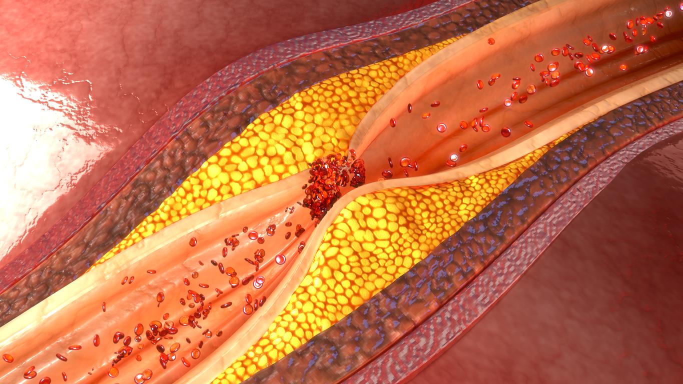
TO?? at Kosair Children’s Hospital in Louisville, heart surgeon Erle Austin was asked to operate on the heart of Roland Lian Cung Bawi, a 14-year-old baby. month whose heart contained blemishes. Without a delicate operation, this little Burmese was doomed to live only a few months with a chronic illness.
Pediatric heart operations are very tricky because they force doctors to work on a very small organ that has not been trained properly. “These structures are difficult to see clearly, even with MRIs (magnetic resonance imaging) and echardiography,” explains surgeon Erle Austin.
To prepare for this sensitive operation, Erle Austin shared 2D images of the diseased heart with other surgeons to seek their advice on how to complete his procedure.
Having only gotten conflicting opinions, Erle Austin decided to call in engineers from the University of Louisville School of Engineering and their 3D printer.
A 3D heart to prepare for the operation
In medicine, 3 D printing is used to create models of human bones, organs, medical devices and prostheses. The surgeon asked specialists to reproduce a model of Roland’s heart with its structure and its flaws.
School principal Tim Gornet and his team managed to reproduce Roland’s heart in 3D, but twice as big as his actual size. “If a picture is worth a thousand words, then a 3-D model is worth 1000 pictures,” said Tim Gornet.
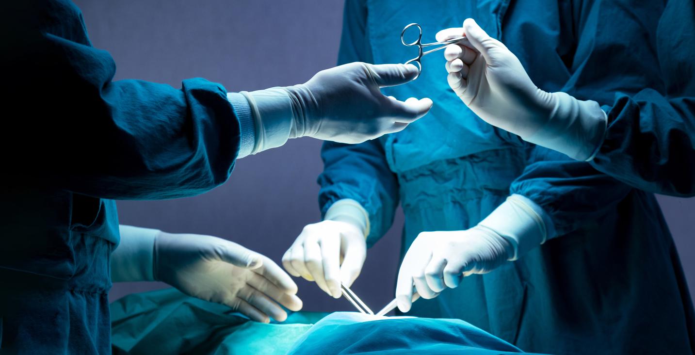
“Once I got this 3D model, I knew exactly what I needed to do and how I could do it,” Erle Austin told the US daily Courier Journal. “Thanks to this 3 D heart, I was able to reduce the exploratory incisions, the operating time and ensure that Roland was born. no need for post-operative follow-ups. It was a huge plus. “
This first operation with a 3D printer, carried out on February 10, was a total success.


