Magnetic resonance elastography, an ultrasound imaging technique, can detect early temporal epilepsy, one of the most treatment-resistant forms.

- Current detection methods for this form of epilepsy fail to visualize epilepsy-induced changes in the brain only after significant damage.
- The results showed a more rigid hippocampus in the group of patients with epilepsy.
- ERM allows earlier detection of the disease, allowing better management.
The hippocampus can serve as a gateway to examine the presence of early temporal lobe epilepsy. Using magnetic resonance elastography, an ultrasound imaging technique usually used to examine the liver, it is possible to detect this pathology by studying the rigidity of the hippocampus. This is the conclusion of a study conducted by American researchers from the Beckman Institute at the University of Illinois (USA), the results of which were published on September 2 in the journal NeuroImage Clinical.
Hit the surface of a pond and watch the ripples
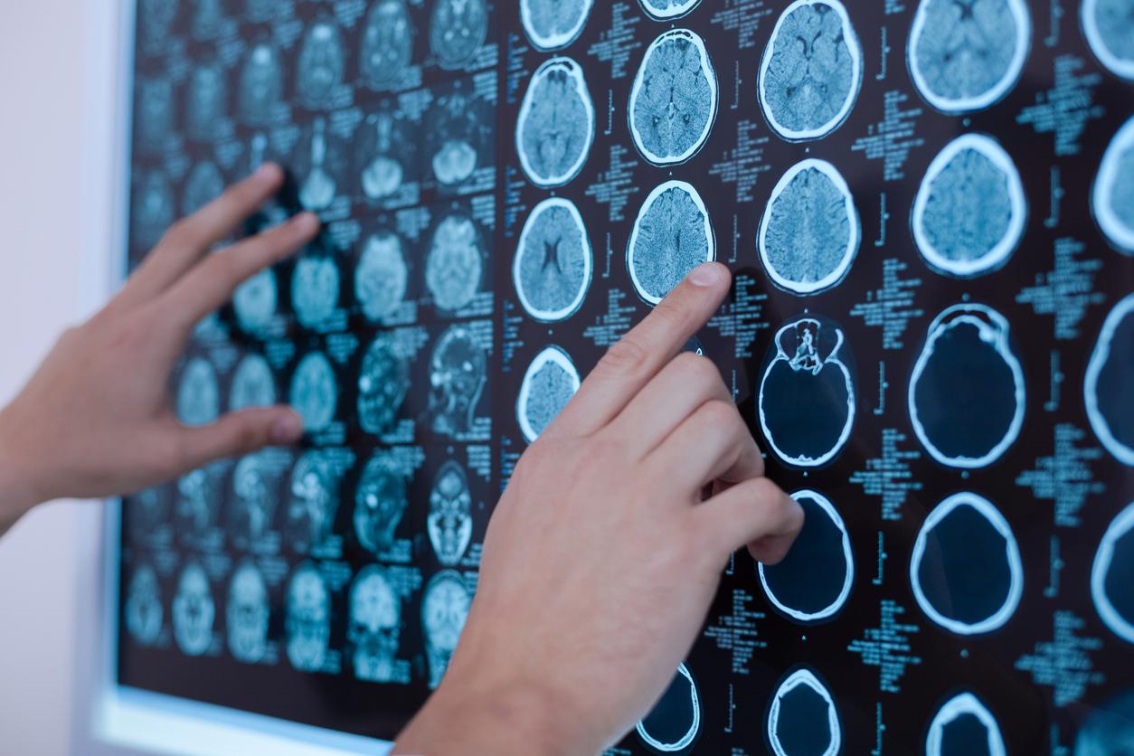
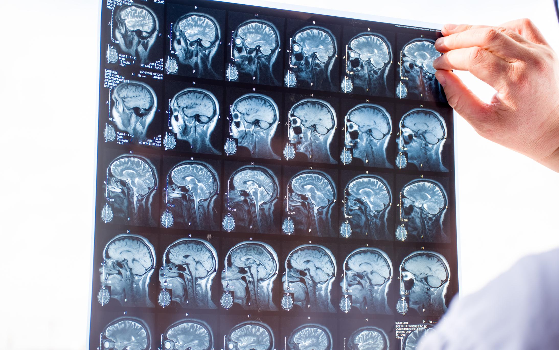
Current detection methods for this form of epilepsy, which include magnetic resonance imaging, only fail to visualize epilepsy-induced changes in the brain after significant damage. This has the consequence that the diagnosis is late and complicates the management of the patient. “Structural changes in the brain, in response to seizures, cause neurons to die and scar tissue to formsays Graham Huesmann, author of the study. By the time we see changes on the MRI, the disease is quite advanced. We wanted to detect these changes earlier using ERM.”
The researchers compared the hippocampus of patients with one of the most common forms of drug-resistant epilepsy, called temporal lobe epilepsy with mesial temporal sclerosis, with that of healthy patients. For this, they used an ERM, a non-invasive technique delivering ultrasound through a small vibrating pillow “sending the vibrations into the changing tissues as the composition and organization of the tissues changeadds Hillary Schwarb, who participated in the study. It’s like hitting the surface of a pond and watching the ripples form. If there is a large rock under the surface, these ripples will move and change.”
Early detection
The results showed a more rigid hippocampus in the group of patients with epilepsy. “The hippocampus is the part of the brain involved in memorysays Brad Sutton, one of the authors of the study. In the early stages of epilepsy, there is a little damage to the structure, which we can detect with ERM.” This early detection is essential, especially since this disease causes very mild symptoms at first. “It starts with a sense of deja vu, which becomes more common as the disease progressesdescribes Graham Huesmann. Eventually, it develops into a drug-resistant form. ERM allows us to detect these changes earlier, allowing us to change the course of treatment.”
Researchers are now focusing on how to optimize the technique and are also looking at other types of epilepsy. “All of our imaging techniques currently depend on examining brain chemistry and static brain imagessays Graham Huesmann. Using ERM to see how the brain moves is an interesting way to tackle this problem. It is also an inexpensive technique and can therefore be used by anyone.”


