Functional brain imaging has made it possible to advance in the understanding of dyslexia and to consider new methods for learning.
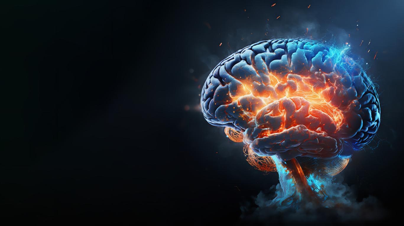
What’s going on in our heads. The question haunts scientists who, every year, on the occasion of Brain Week (March 10-16), combine their data and their research. This meeting is an opportunity to measure the progress made in neuroscience. Functional brain imaging (ICF) has made it possible, for example, to deliver information on the brain in activity within a few years. It thus becomes possible to observe disturbances in cerebral functioning and to understand, for example, what is happening in the brain of a dyslexic.
The ICF does not necessarily provide insight into dyslexia. On the other hand, it makes it possible to obtain cerebral correlates on what experimental psychology has already made it possible to understand. “We have now acquired a certain amount of knowledge about the development of the brain. Imagery can provide answers if we explore a particular hypothesis, for the moment on a mainly descriptive process, ”explains Franck Ramus, research director at the Laboratory of Cognitive and Psycholinguistic Sciences at the Ecole normale supérieure. “In dyslexia, for example, it has been observed that certain areas of the left brain are not activated in the same way as in a non-dyslexic brain.”
Dyslexia is characterized by difficulty in matching graphemes (letters and groups of letters) with phonemes (speech sounds). The regions affected by a change in activity correspond well to these observations since they are located in the occipito-temporal lobe, through which the visual information enters, the temporo-parietal lobe, which provides access to the lexicon and the lobe. frontal which allows the pronunciation of the word. Functional brain imaging also found that in some dyslexics homologous areas of the right brain were overactivated during reading, as if to compensate for left brain defects.
Four associated genes
“To understand these data, we then had to question what is fundamentally different in the brain of a dyslexic, what happened differently during his development,” recalls Franck Ramus. The great advance today is to have been able to go back to the genetic level for cognitive learning and to have found four genes associated with dyslexia, which we now know are involved in neuronal migration. Genetics here confirm hypotheses established by brain imaging since the images reveal unusual concentrations of neurons in the left brain of dyslexics, which in fact correspond to neurons that have not migrated in a normal way (see opposite ).
Brain imaging can now confirm the effects on the brain of many methods of rehabilitation of dyslexics. It is thus possible to improve the situation of dyslexic children and teach them to read by playing on the natural plasticity of the brain. More simply, if we provide them with effective rehabilitation such as good speech therapy, researchers have observed that non-activated areas of the brain start to function again. “Imaging studies show that normalizations may be linked to a certain type of rehabilitation,” says Franck Ramus. “New studies could also help determine subtypes of dyslexia and what types of therapy might work best.”
Evaluate learning
At present, neurosciences have made it possible to provide a set of answers on the origin of dyslexia and the way in which it manifests itself. Neuroscientists can now offer rehabilitation and learning approaches based on this knowledge, but it is difficult to transfer this information to teaching teams. Inserm’s collective expertise on learning disorders, published in early 2007, highlights the current consensus of scientists on this pathology but indeed establishes that screening, diagnosis and treatment could benefit from a rapprochement between neuroscientists, learning disability management teams and teaching teams.
“The better we know the brain, the more we will be able, for example, to suggest methods for better learning. These methods will have to be scientifically evaluated before they can be recommended and disseminated ”, specifies Franck Ramus. In France, no learning method has ever been rigorously evaluated, with control groups. For the researcher, the main difficulty lies in the fact that educational specialists do not have the scientific culture that would allow them to use neuroscience data to design new methods but also to evaluate them.
The evaluation is therefore carried out empirically. “A link in the chain is really missing and today, I at least try to educate the teaching world on what it is possible to do, at their level, because the methods at their disposal do not take the news into account. brain data. If it were already possible to ensure that teachers use one method among those which are correct, we could reduce the number of children who develop dyslexia, ”insists Franck Ramus. And, to prepare the methods of the future, he also plans to train doctoral students who would have this double view on neurosciences and pedagogy.
.


