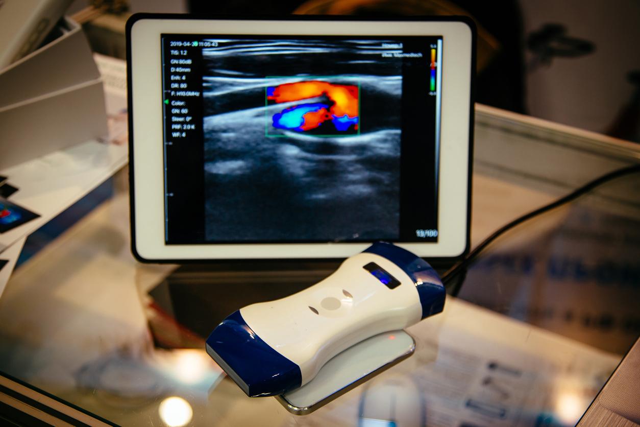
Doppler ultrasound(more commonly known as Doppler ultrasound) of the lower limbs is an exam that allows you to get a picture of the vessels and to check the flow of blood in the arteries of the legs and feet. This examination combines two techniques: ultrasound which allows to study the morphology of the vessels and to visualize the veins, and the Doppler which makes it possible to visualize the way in which the blood flows in the veins and to measure its flow speed. . This is why the doctor prescribes a Doppler ultrasound to be done within 24 hours, when he suspects superficial or deep phlebitis.
Echo-doppler: how is it going?
This exam is performed without any special preparation: it is completely painless and lasts an average of 30 minutes. You are lying on an examination table. The angiologist applies a gel to the skin in the arteries of the lower limbs, to facilitate the transmission of ultrasound. He then places the ultrasound probe in contact with the skin and moves it with light pressure. Ultrasound echoes are recorded by a computer which converts them into a video image. The doctor saves the images on which he wishes to measure parameters, for example the width of an artery or the thickness of its wall.
The doctor will locate on the images which are the dilated areas of the veins and will be able to locate the diseased parts with precision. The entire venous system of the lower limbs is accessible by color Doppler echo. Also, when there is a clot (called a thrombus) in a vein, it is perfectly visible.
If the Doppler echo rules out the diagnosis of deep vein thrombosis (or phlebitis), it can also highlight the cause of the abnormally swollen leg (for example the presence of a cyst behind the knee or simply a insufficient venous circulation).


