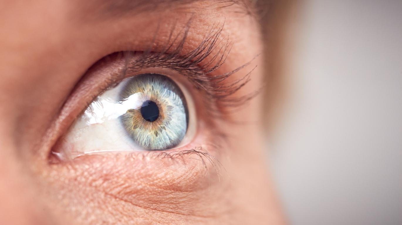
A new non-invasive imaging system has just been developed to facilitate the diagnosis and treatment of dry eye responsible for irritation and visual disturbances.

Suffering from dry eye means often having irritated eyes and especially vision problems. Who cause often a irritation and a vision trouble. A A study carried out at the AdOM laboratory (Advanced Optical Methods) announces a new method for diagnosing and treating this disease.
What is dry eyes?
Dry eye is a disease caused by tear film instability. Our eyes are moistened by the tear film which protects, cleans and hydrates them. Thanks to the blinking of the eyes, this film is renewed and evenly distributed on the surface of the eye. In people with dry eye, the tear film does not moisten the eyes properly, either because the tear fluid is produced in insufficient quantity or because its composition is changed. This therefore leads to unpleasant symptoms that can affect our performance and well-being.
Today, most cases of dry eye are diagnosed using a questionnaire, which is of low reliability. Subjective symptoms are often poorly or badly correlated with objective signs. The tear film being the first system crossed by light, the optical quality of the eye depends strongly on the homogeneity of the tear film. Objective methods of tear film examination usually cannot keep up with changing dynamics and therefore cannot be used to identify the cause of the disease.
Improving diagnosis through imaging
“Up to 60% of visits to the ophthalmologist are due to dry eye, which shows the need for a non-invasive and very precise device to carry out the diagnosis in the city,” said Yoel Arieli, head of the AdOM (Advanced Optical Methods Ltd.) research team in Israel.
In the review Applied Optics, the researchers described the device’s ability to perform measurements over a large field of view in seconds. The image can acquire rapid and consistent measurements of human eyes, even when blinking. The new instrument uses eye-safe halogen light to illuminate the eye, then analyzes the full spectrum of light reflection in time and space. These spectral measurements are used to reconstruct the structures found in the front of the eye, allowing precise measurement of the inner layers of the tear film, particularly the aqueous sublayer. This sublayer plays an important role in dry eye, but has been difficult to analyze with other methods.
“Tear film imaging is the first device that can be used in the ophthalmologist to reflect the tear film and distinguish its inner layers with nanometer resolution, those measures are carried out automatically in only 40 seconds. says Yoel Arieli.
A non-invasive device that would present no risk
After to have demonstrated a resolution of 2.2 nanometers on a mock tear film, the researchers have tested the capacity of the tool at take from measures on the eye human without intervention during than the patient winked. “ The device at worked of way impressive and born present any risk by that he is not not invasive and uses a source luminous simple », at declared the Dr. Arieli. “ He at no only measure the movie tear of way consistent, there understood the blinks all the seconds, corn the measures to are Good correlated with others techniques of diagnostic partially invasive. »
Tear film imaging has been used in two clinical studies in Israel and Canada. The dry eye diagnostic review has been completed with the device and treatments can now be accurately assessed. However, the researchers affirm than of future studies could to help better prescribe treatments, to improve the results surgical and lead to an adjustment from lentils of contact. They are considering also to use the device in from studies more vast with from groups more various of patients for to define from levels for the eyes healthy and the people suffering from dry eye.
.


