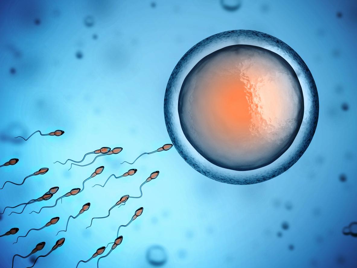
Thanks to 3D imaging, researchers have succeeded in showing the presence in human embryos of reptile muscles which disappear after birth. These anatomical structures have not existed in mammals for 250 million years.
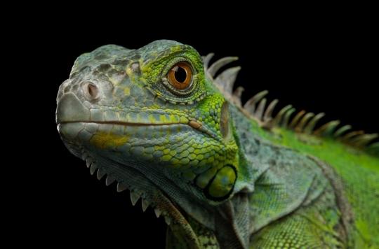
Since Darwin revolutionized the history of biology with the publication of his book The Origin of Species in 1859, many scientists assure that the punctual appearance of atavistic structures (anatomical structures lost during the evolution of a certain group of organisms that can be present in their embryos or reappear in adults as variations or anomalies) proves species change over time.
While small tail-like structures appear temporarily in human embryos and the rest are preserved as coccyxes, several researchers believe that atavistic muscle and bone can also be seen in human embryos. Today, thanks to a new technology providing high-quality 3D images of embryos and fetuses, American researchers have succeeded in identifying them. The results of their study were published on 1eroctober in the magazine Development.
Dr Rui Diogo of Howard University (USA) and his colleagues used 3D images to produce the first detailed analysis of the development of human muscles and legs. This unprecedented resolution revealed the transient presence of several of these atavistic muscles. In the hand and foot, out of thirty muscles formed at about 7 weeks gestation, one-third will fuse or disappear at about 13 weeks gestation. This decrease corresponds to what happened in evolution more than 250 million years ago, when certain reptiles began to transform into mammals.
“See human development in much greater detail”
These muscles allow the fingers and toes to move in almost any direction. Only one of them persists in human embryos: that of the thumb of the hand, the only finger that opposes the palm and allows us to grab objects. It is this particularity, which our species shares with other primates, which allowed the manufacture of the first tools or the emergence of art.
“We used to understand the early development of fish, frogs, chickens and mice better than in our own species, but these new techniques allow us to see human development in much greater detail. is that we have observed various muscles that have never been described in human prenatal development, and some of these atavistic muscles have been observed even in 11.5 week old fetuses, which is very late for developmental atavisms “, comments Doctor Diogo.
New perspectives
This discovery therefore deconstructs the myth that in our evolution and prenatal development, we become more complex with more anatomical structures such as muscles continuously formed by the splitting of anterior muscles. These results offer new perspectives on the evolution of our arms and legs since those of our ancestors but also on the variations and certain human pathologies.
Indeed, “it is interesting to note that certain atavistic muscles are rarely found in adults, either in the form of anatomical variations without noticeable effect for the healthy individual, or as a result of congenital malformations”, explains Diego. And to conclude: “This reinforces the idea that variations and muscular pathologies may be related to delayed or arrested embryonic development, in this case possibly delayed or decreased muscle apoptosis, and helps explain why these muscles are sometimes present in adults. It is a fascinating and powerful example of the evolution taking place.”
.


