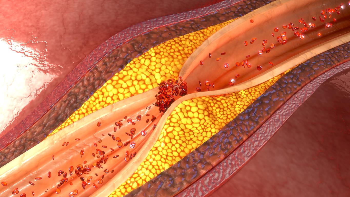
A new non-invasive technique would detect inflammation in the arteries, thus detecting a heart disease in individuals at greatest risk, according to the results of thestudy published in the medical journal Science Translational Medicine.
Today, doctors are detecting a heart disease using a CT scan to identify arteries that are severely damaged by the build-up of plaques that restrict blood flow. But this technique is far from perfect.
Often screening is done while the patient is in critical condition. In fact, the narrowness of the arteries is not always the signal of an approaching heart attack, unlike inflammation.
“Until now, there was no way to detect inflammation in the coronary arteries,” said Keith Channon, professor of cardiovascular medicine at the University of Oxford.
A new test to identify inflammation of the arteries
Researchers at the University of Oxford in the UK have developed a process that analyzes changes in the fatty tissue that surrounds the arteries, known as perivascular fat, from images from a CT scan. This fat becomes more watery and less fatty when it is near an inflamed artery.
Scientists also found that they could track changes in perivascular fat over time, allowing doctors to spot early signs of heart disease that could be prevented with taking statins lowering the rate of cholesterol.
While the results of this study are encouraging, further study is needed to confirm that this approach can predict heart attacks and save lives.
“I am optimistic and I believe that this technique will allow us to predict future cardiovascular accidents but we have to confirm the results in larger studies, “said Charalambos Antoniades.
Read also:
Broken heart syndrome: long-term sequelae
Vegetables to protect the arteries and the heart
Breastfeeding would protect women from cardiovascular disease


