


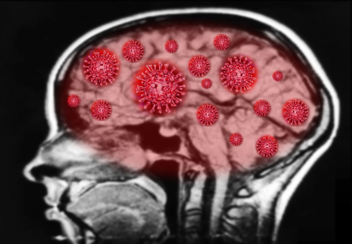
Cerebral, immune and metabolic abnormalities are thought to be linked to myalgic encephalomyelitis.

- Exhaustion is thought to be caused by dysfunction in brain regions controlling the motor cortex, such as the temporoparietal junction, which causes fatigue by disrupting the way the brain decides how much effort to exert.
- Adults with chronic fatigue syndrome had low levels of catecholamines, which are linked to worse motor performance, exercise-related behaviors and cognitive symptoms.
- Differences between men and women were observed in gene expression patterns, immune cell populations, and metabolic markers.
Crushing fatigue, discomfort after exercise, unrefreshing sleep… Chronic fatigue syndrome, also known as “myalgic encephalomyelitis”, has nothing to do with temporary losses of vitality. It’s about a “disabling disease whose clinical phenotype is poorly defined, the pathophysiology is unknown and for which there is no modifying treatment”, according to researchers from the city of Bethesda in Maryland (United States).
Chronic fatigue syndrome: 17 patients underwent medical evaluation
In a recent study, they determined all the observable characteristics of adults with myalgic encephalomyelitis/chronic fatigue syndrome (ME/CFS). For this, the scientists recruited 21 healthy adults and 17 patients suffering from this disease, which developed after a viral or bacterial infection, for five years. Volunteers had to make risk-based decisions about physical exertion. This allowed the team to assess the cognitive aspects of fatigue, that is, how a person decides how much effort to exert when given a choice.
Additionally, participants underwent a multi-day ME/CFS medical screening and evaluation and underwent extensive testing, including clinical examinations, physical and cognitive performance tests, autonomic function tests, skin and muscle biopsies, and advanced blood and spinal fluid analyses. People also spent time in metabolic chambers where, under controlled conditions, their diet, energy consumption, metabolism, sleep patterns and gut microbiota were assessed. During a second visit, they performed a cardiopulmonary exercise test to measure the body’s response to exercise.
Fatigue comes from “a mismatch between what a person thinks they can accomplish and how their body is performing”
According to the results, published in the journal Nature Communications, while no signs of muscle fatigue were observed, participants affected by ME/CFS experienced difficulties in the effort choice task and in maintaining effort. The motor cortex, a region of the brain responsible for telling the body to move, also remained abnormally active during the strenuous tasks.
According to the researchers, this suggests that fatigue in this disease could be caused by dysfunction in brain regions controlling the motor cortex, such as the temporoparietal junction (TPJ), which can cause fatigue by disrupting the way whose brain decides how much effort to make. “So, rather than physical exhaustion or a lack of motivation, fatigue may come from a mismatch between what a person thinks they can accomplish and how their body is performing,” said Brian Walittmain author of the work.
Low levels of catecholamines are linked to poorer motor performance
Cerebrospinal fluid analyzes showed abnormally low levels of catecholamines and other molecules, which help regulate the nervous system in sick people compared to healthy volunteers. These low levels have been associated with worsening motor performance, exercise-related behaviors, and cognitive symptoms. “We believe that immune activation affects the brain in diverse ways, causing biochemical changes and downstream effects, such as motor, autonomic, and cardiorespiratory dysfunction,” explained Avindra Nath, co-author of the study.
Another finding: Immune testing revealed that the ME/CFS group had higher levels of naive B cells and lower levels of switched memory B cells, which help the immune system fight pathogens in the blood. .

Marked differences between men and women with ME/CFS
The data revealed differences between men and women in gene expression patterns, immune cell populations and metabolic markers. Men showed impaired T cell activation, as well as markers of innate immunity, while women showed abnormal growth patterns of B cells and white blood cells. Men and women also had distinct markers of inflammation.
“Given the immune differences between men and women in ME/CFS, the results may open new avenues of research that could lead to a better understanding of other chronic diseases associated with infections. (…) Researchers can now verify whether these data apply to a larger group of patients and make progress in identifying treatments that target the main drivers of the disease”, the scientists concluded.


