


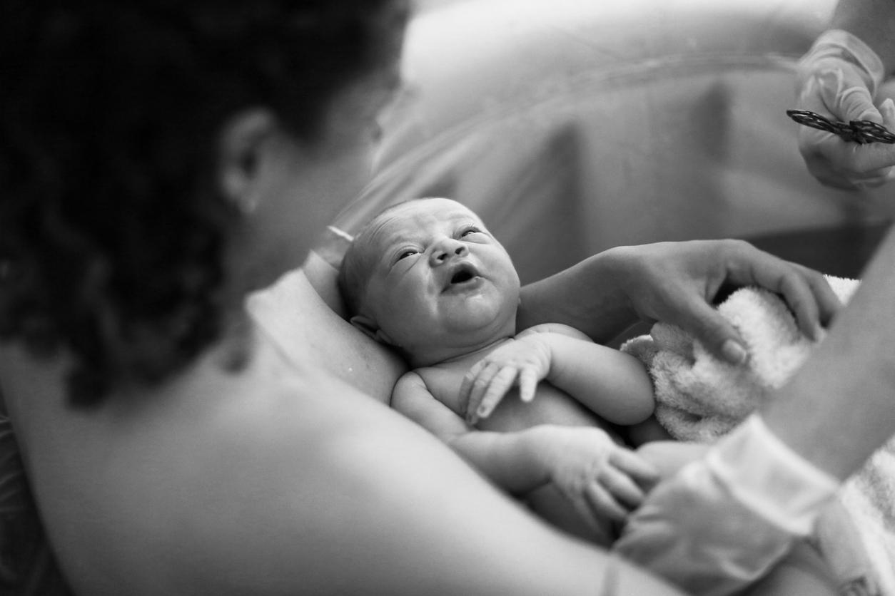
For the first time, a French team of researchers has managed to capture 3D images of infants born vaginally and thus observe the cranial deformation they undergo during childbirth.

Who has never noticed in a newborn the elongated shape of his skull? This phenomenon is well known to doctors. Called the “casting” of the fetal head, this change in cranial shape occurs during the second stage of vaginal birth, when the baby leaves the uterus and begins to move into the pelvic cavity.
But so far, the details of cranial deformation, especially when these changes occurred, have remained unclear to researchers. New work carried out by the team of Dr Olivier Ami, from Clermont-Ferrand University Hospital, and published on May 15 in the journal PLOS Oneprovide new answers.
Using magnetic resonance imaging (MRI), researchers captured 3D images showing how infants’ brains and skulls twist as they move through the pelvic canal.
A “sugar loaf” skull
After obtaining approval from an ethics committee, the medical team performed two MRIs on seven women whose pregnancies had gone off without a hitch. A first session took place a few weeks during childbirth, the second during the second phase of labor, when the newborn’s head enters the cervix. “The goal was to document what is happening biomechanically in the baby’s skull and brain,” explains the Figaro obstetrician-gynecologist Olivier Ami.
Analysis of the images revealed that the newborns “showed a deformed head with overlapping cranial sutures”. “The modification of the fetal head was systematically observed when the head was engaged between the superior strait and the superior edge of the pubic bone”, detail the researchers in their publication. “For the 7 children, the skull adopted the same so-called ‘sugar loaf’ shape: their skull was deformed upwards by compressing laterally”, explains Olivier Ami.
After birth, five of the newborns saw the skull and brain shapes return to their prenatal state. However, the changes persisted in two of the infants. The study does not say whether the situation subsequently improved.
Two of the three infants with the highest degree of fetal head molding delivered by emergency caesarean section. The third, however, was delivered vaginally with minimal expulsive effort.
Very high cranial pressure
These new findings suggest that infants experience more cranial stress during birth than previously thought. According to the researchers, this cranial pressure could explain the occurrence of asymptomatic brain and retinal bleeding observed in many newborns after vaginal delivery. “During vaginal birth, the shape of the fetal brain undergoes a greater or lesser deformation depending on the degree of overlapping of the bones of the skull. The molding of the fetal skull is no longer visible in most newborns after birth Some skulls accept the deformation and allow easy expulsion, while others do not deform easily,” explains the doctor.
This is the second study looking at cranial deformities in babies during childbirth. “The first was done in Germany a few years ago but it only involved one woman and used a 2-dimensional MRI,” says Dr Ami.
.


