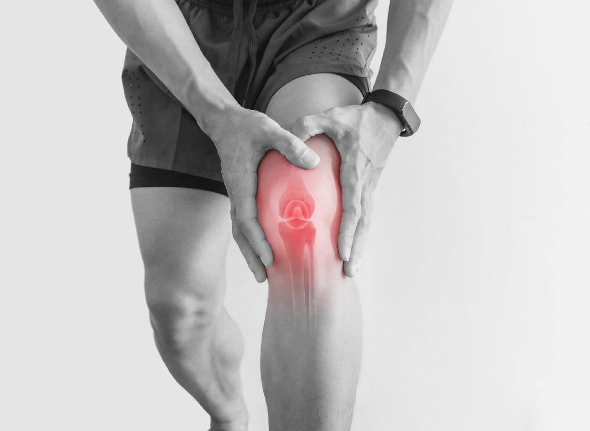
In the United States, a professor of physiotherapy from the University of Montana published a clinical practice guideline on Sunday to improve the management of patients suffering from patellofemoral pain syndrome (PFPS), better known as “knee of the runner”.

The pain syndrome patellofemoral (SDFP), commonly known as “runner’s knee”, is one of the most common causes of anterior knee pain in adolescents and adults. Every year, it affects a quarter of the world’s population and twice as many women. Unfortunately, despite its frequency, this disorder is still poorly understood. Also its diagnosis is complicated and its management difficult.
This is why Richard Willy, professor of physiotherapy at the University of Montana (USA) published a Clinical Practice Guideline to improve standardization of patient care while providing reimbursement guidelines to insurance companies. Its recommendations were published on Sunday 1erSeptember in the Journal of Orthopedic & Sports Physical Therapythe official scientific journal of the Academy of Orthopedic Physical Therapy.
According to him, exercise therapy, namely hip and knee strengthening treatments prescribed by a physiotherapist, is the best approach for the recovery of patients.
Specializing in one sport is more risky
Recalling in passing that young athletes who specialize in a single sport run a 28% greater risk of suffering from SDFP, Willy recommends to avoid it an exercise program to gradually increase activities such as running or walking. . Maximizing leg strength, especially the thigh muscles, further reduces risk. Finally, pain does not always mean that the knee is damaged, he explains.
“While it may be tempting to seek quick fixes for knee pain relief, there is no evidence that non-active treatments alone, such as electrical stimulation, lumbar manipulation, ultrasound, or dry needles, help sufferers. of SDFP,” he also said. And to conclude: “People with PDPS should consult a clinician who uses exercise therapy to treat this injury.”
SDFP results from poor routing of the patella (kneecap) during mobilization of the knee, which then leads to excessive compression on the patellar facets. At the present time, in France, its diagnosis is made mainly in relation to the account of the patient’s history and to the clinical examination of the knee and the whole of the lower limb. An MRI is sometimes done. When the diagnosis is made, the treatment is often mainly focused on rehabilitation with targeted and personalized physiotherapy, as Willy advises. Surgical treatment is reserved for cases with a causal structural abnormality.

.


