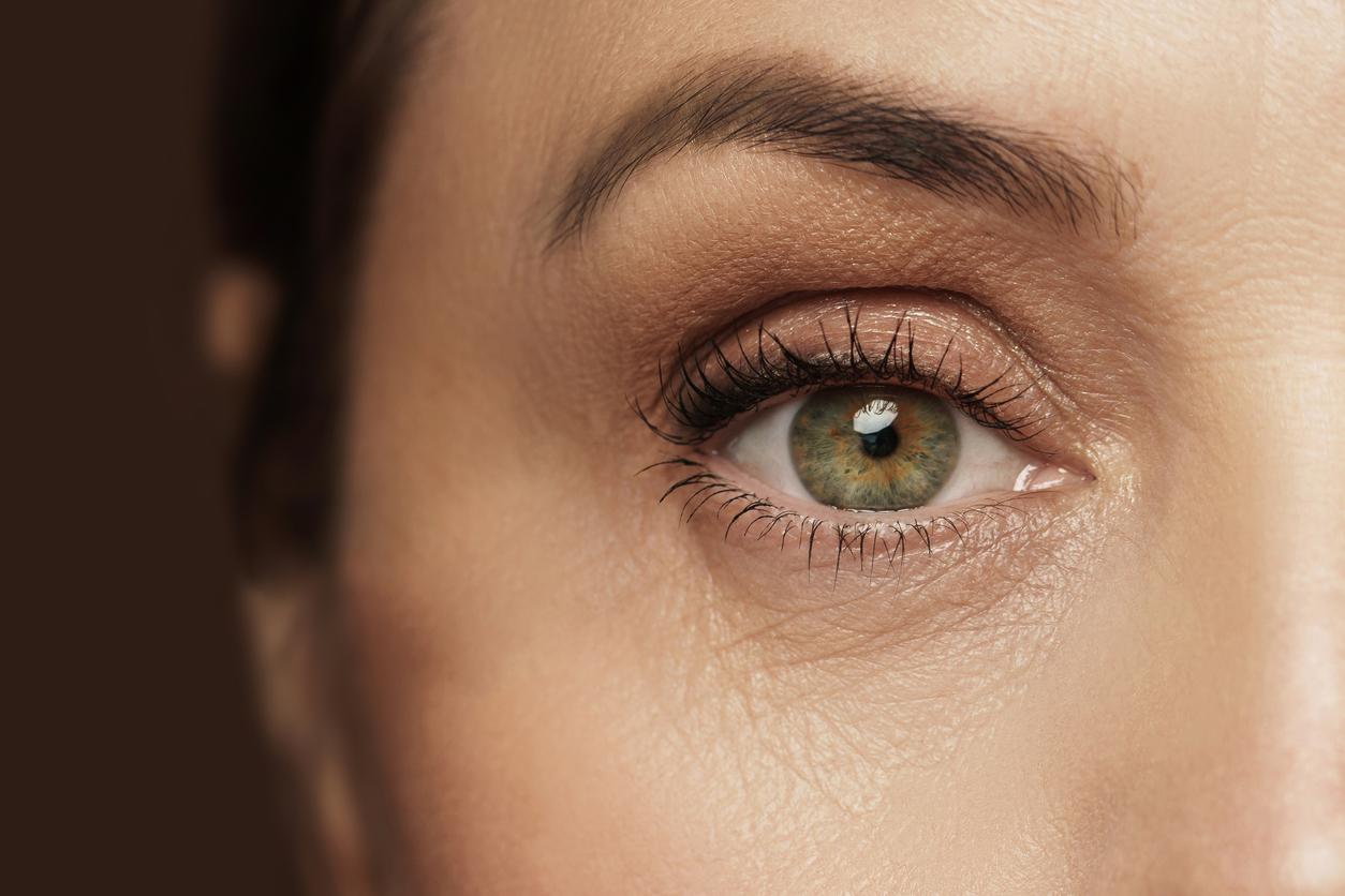
Thanks to new technologies, researchers have been able to explore new areas of eyelash structure in order to understand the origin of certain eye diseases.
-1573729658.jpg)
In addition to enhancing the looks, eyelashes play an important role in our vision. Their main function is to prevent dust and sweat from getting into our eyes. In addition, like blinds, eyelashes also serve to filter some of the light, especially when we squint or close our eyes. However, any ciliary abnormality can be a potential cause of retinal degeneration and blindness. Until now, traditional imaging technologies have limited the examination of ciliary structures, but US researchers from Baylor College of Medicine and McGovern Medical School (Texas) have conducted a new study that allows us to see the eyelashes, to see what might be associated with vision loss. The study was published in the American journal Proceedings of the National Academy of Sciences.
In their study, the researchers developed and applied new imaging methods that provide a high-resolution view of the structure of normal and diseased retinal cilia. This new technique has made it possible to discover new compartments and new proteins in the eyelashes that could be involved in retinal diseases. Ultimately, this research will pave the way for an in-depth study of the specific structural defects of the eyelashes that lead to blindness, while facilitating the development of new treatments.
A milestone
According to Dr. Theodore Wensel, professor and chair of chemistry in Baylor’s biochemistry department, this is an important discovery. “There is a large group of diseases called ciliopathies, which are caused by genetic defects in the structural components of the eyelashes. One of the most common symptoms of ciliopathies is retinal degeneration and blindness, such as seen in retina pigmentosa and Bardet-Biedl syndrome,” he explains. Bardet-Biedl syndrome is a genetic disorder that affects many parts of the body, but particularly the eyes. In particular, it causes a narrowing of the visual field and loss of vision at night.
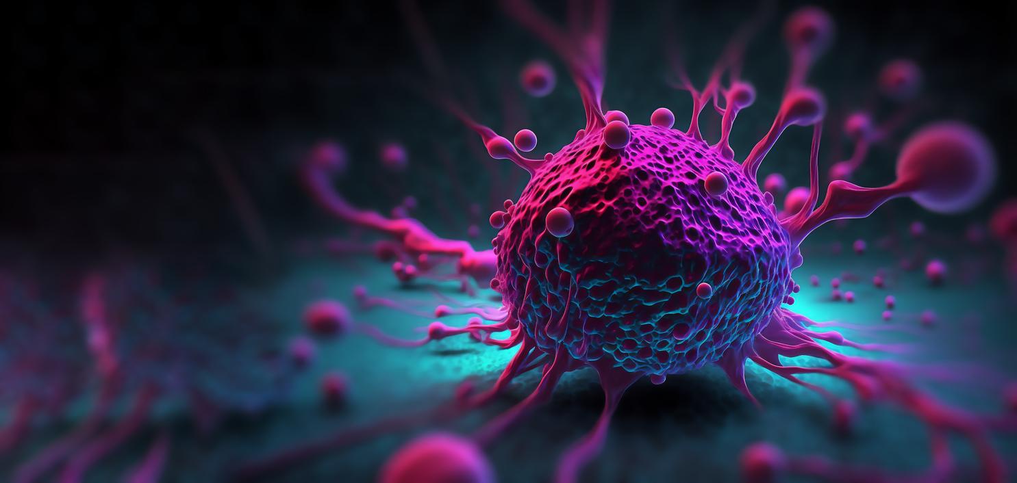
Previous attempts to better understand how genetic defects associated with retinal ciliopathies have been delayed by minute eyelash size. Traditional methods could not reveal the internal structure of hair cells. This time around, the researchers developed cryo-electron tomography (to make “sections” of the eyelashes) and super-resolution stochastic optical reconstruction microscopy (STORM), which gave a deep nanoscale view of internal structures. Eyelash. When the researchers used these methods to analyze the cilia, they saw structures that had never been seen before. In diseased retinas, these high-resolution techniques have made it possible to locate proteins associated with disease more precisely.
Ciliopathy and ocular malformations
The new techniques have considerably improved the results. In elaborating on this subject, Theodore Wensel thought that a particular protein encoded by a disease gene was located in the eyelash. “Now we can say that it is located in the center of the axon, which is a tiny structure located in the center of the cilium, or in another of the newly discovered compartments.” Additionally, the researchers discovered genetic defects that changed the location of certain proteins in the structure. This discovery may contribute to the development of new therapies.
Ciliopathy is the name given to a group of disorders related to mutations in the hair proteins causing abnormalities in the cilia. Such abnormalities in eyelash function have been observed in several disorders, including retinitis pigmentosa, which is the most common form of retinal degeneration worldwide. It can cause conditions such as night blindness, tubular vision and blindness.
.


