Researchers have managed to “tag” CAR-T cells reintroduced into patients with molecular markers to better understand their functioning and monitor their activity against cancer cells.

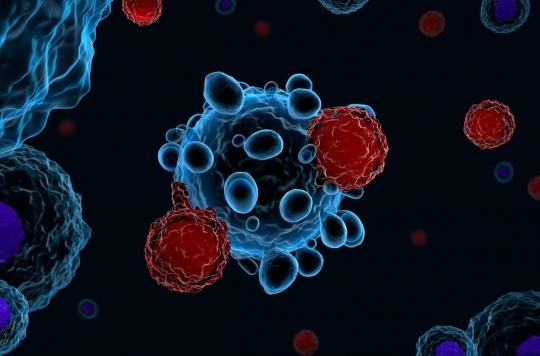
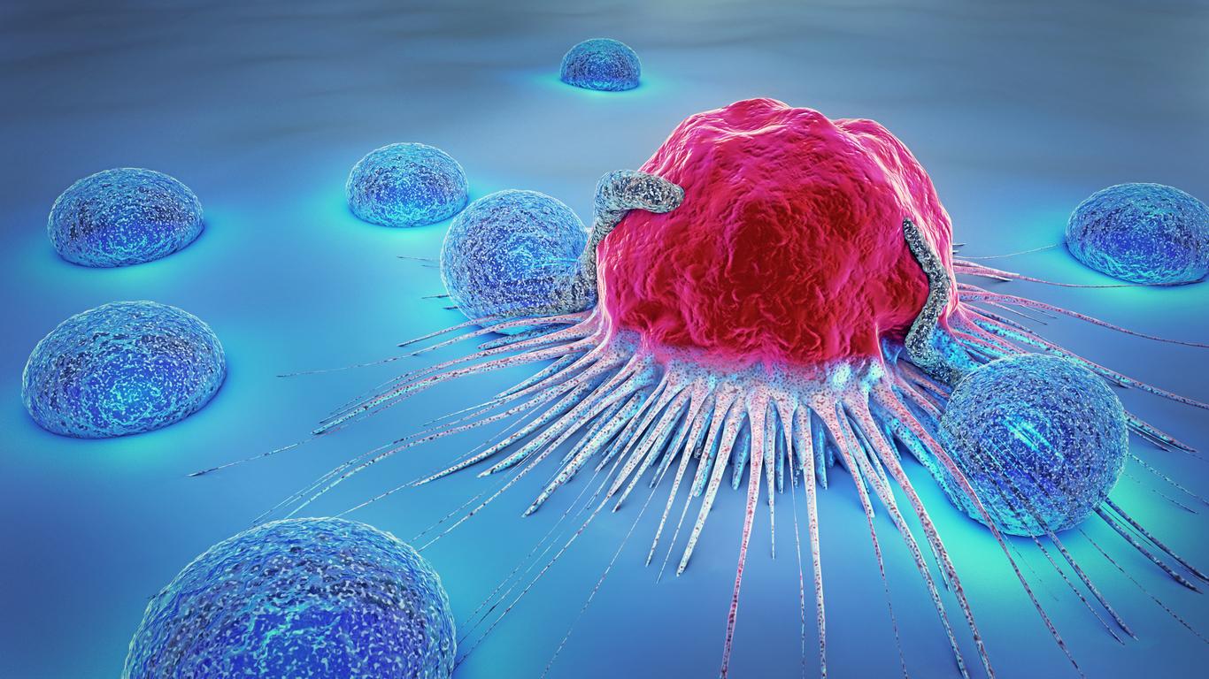
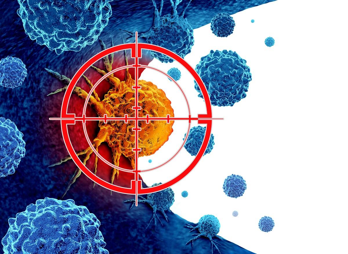
In recent years, immunotherapy based on CAR-T cells has revolutionized clinical treatment against severe forms of cancer. This technique involves taking T cells from the patient’s blood and sending them to a laboratory. Using cell engineering technologies, researchers modify in vitro the genetic material of T lymphocytes in order to endow them with a specific receptor which acts as a radar to recognize and attack the cells expressing the target antigen. The number of T lymphocytes thus “reprogrammed” is multiplied in the laboratory, then the modified cells are reinjected into the patient.
Monitoring CAR-T cells with a molecular marker
But so far, researchers have come up against an obstacle: how do you know where the CAR-T cells go in the body once they have been reinjected into the patient? “Currently, the only way to know if gene or cell therapy is still present in the body is to regularly biopsy tumors or draw blood, which provide very crude measures of therapy,” says Professor Mark Sellmyer, assistant professor of radiology and researcher at the Perelman School of Medicine at the University of Pennsylvania.
With his team, he found how to track reprogrammed T lymphocytes in the body: they genetically modified them using molecular markers visible using position emission tomography (PET-scan). The results of this study are published in the journal Molecular Therapy.
For their work, the researchers genetically marked the CAR-T cells with a bacterial protein eDHFR (called “PET reporter gene”), which were then injected into mice. These then underwent an injection of trimethoprim, an antibiotic molecule used here as a contrast product: the CAR-T cells then lit up, which allowed the researchers to follow them in real time using the PET scan. And because the cells’ DNA was genetically modified, once the car-T cells multiplied, the new cells could also be tracked by the researchers.
A new step in immunotherapy
Images collected by the researchers showed that after seven days, T cells began to accumulate in the rodents’ spleens, and after thirteen days they had begun to target antigen-positive tumours. For the authors of the study, this suggests that there are early and late anchoring “ports” for CAR-T cells in the body.
“Using our technology, clinicians would be able to see, quantitatively, the number and location of CAR-T cells that have survived in the body over time, which is an indicator of durability and health. potential efficacy of the treatment”, continues Professor Sellmyer. “Imaging CAR-T cells will also make it easier for researchers to test and modify therapies for different types of diseases in the research setting.”
Now, the researchers plan to test this technique in a clinical trial on humans. “The hope for the future is that many gene or cell therapies, like CAR-T, will be labeled and tracked in the body,” concludes the study author.
.


