Patients diagnosed with angina are better off having an MRI than an invasive angiogram.

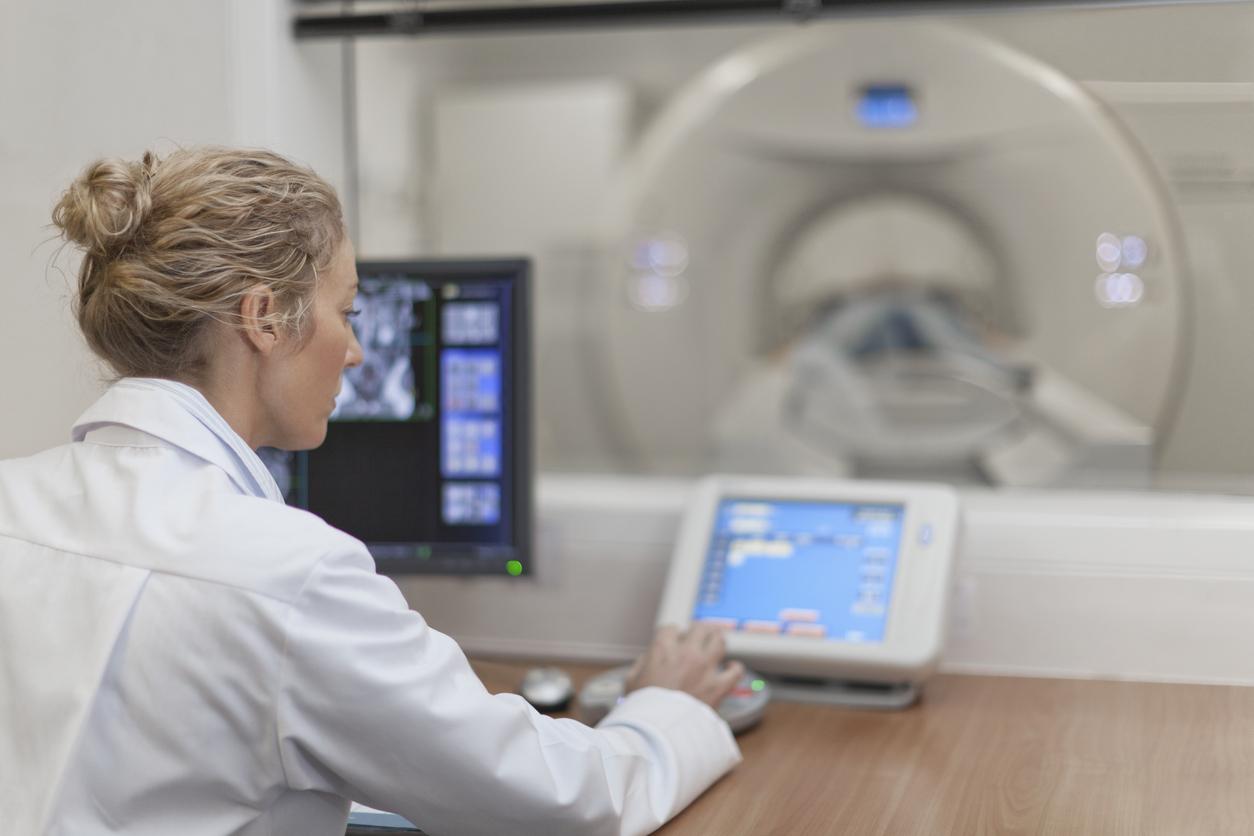
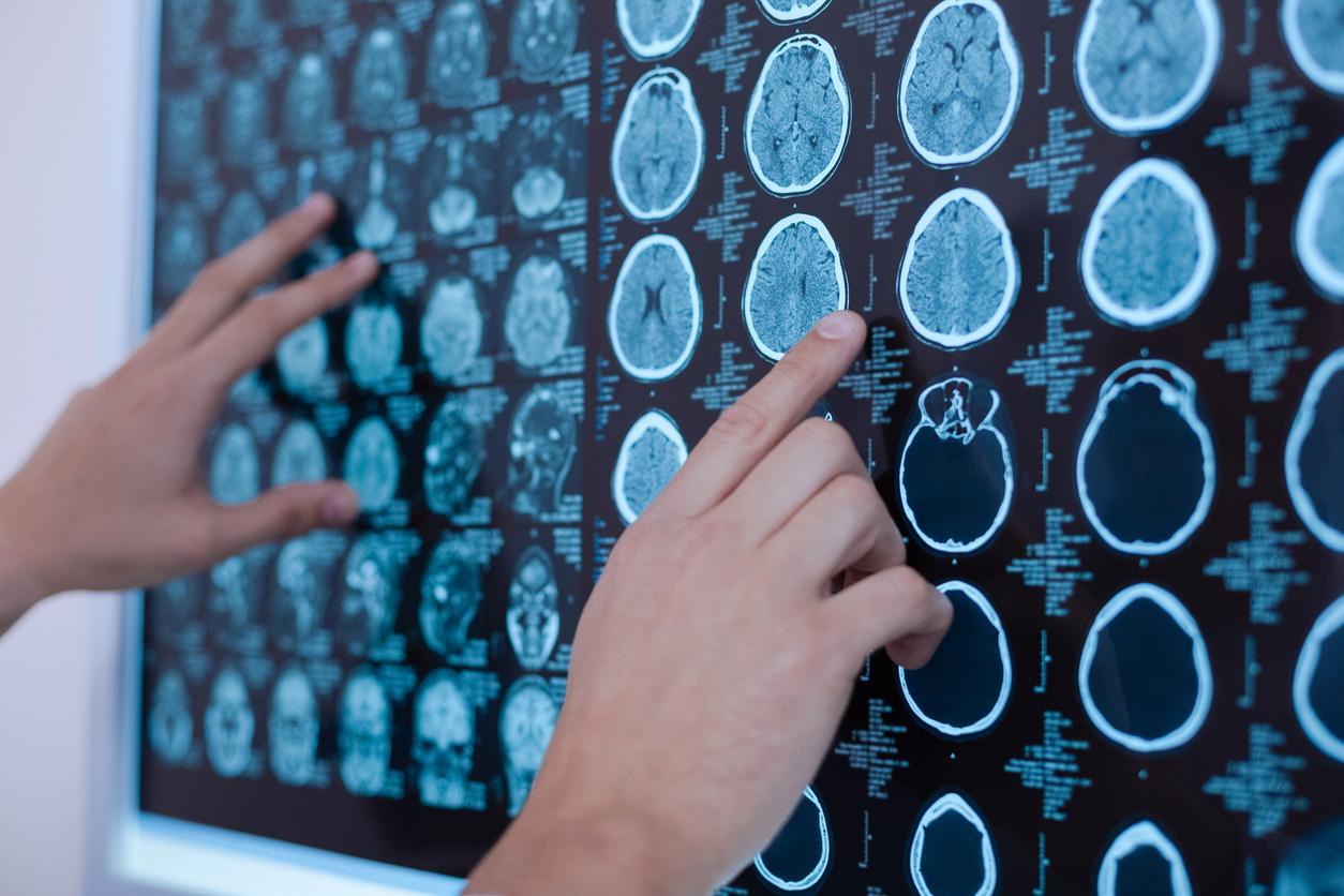
If you suffer from angina pectoris, having an MRI proves effective and non-invasive, in particular avoiding hospitalizations. These are the results of the MR-INFORM trial. The experiment aimed to find out if magnetic resonance imaging (MRI) could be used to guide treatment decisions for patients with angina, rather than performing a more invasive intervention.
Multiple hospital visits
Angina pectoris is chest pain caused by reduced blood flow to the heart muscles. This disease is a warning sign of a risk of heart attack or stroke.
Currently, patients who are diagnosed with angina should undergo invasive angiography, a procedure that involves taking x-rays of the patient’s arteries over multiple hospital visits, and staying overnight. . If the condition is severe, patients should undergo a procedure to improve blood flow to the heart, called “revascularization”.
The MR-INFORM trial involved 918 patients with angina pectoris, who were divided into two groups. One of them had the standard invasive angiogram. The other underwent a 40-minute MRI to decide whether to send the patient for invasive angiography.
Personalize the treatment of angina pectoris
The two groups gave similar results concerning the health of the patients, less than 4% of the patients having had heart problems the following year. However, the group whose treatment was dictated by MRI underwent far fewer interventions, and only 40% of these patients had recourse to invasive angiography. 36% of the MRI group underwent revascularization, compared to 45% in the other group.
For Professor Eike Nagel, cardiologist and director of research: “personalizing the treatment of angina pectoris means that we will be able to target the most invasive treatments only on patients who really need them”.

.


