Coupled with a marker, llama antibodies could early identify lesions linked to Alzheimer’s disease.

One of the challenges of Alzheimer’s disease is the ability of doctors to detect brain damage as early as possible. A team from the Institut Pasteur, in collaboration with Inserm, CNRS, CEA, Pierre and Marie Curie and Paris Descartes universities and the Roche group, has developed a new technique to highlight these lesions in patients. with Alzheimer’s disease, thanks to llama antibodies. They published their results in the journal Journal of controlled release.
As the early manifestations of Alzheimer’s are not always evident in behavior, the first lesions go unnoticed, as conventional imaging does not allow them to be detected. “Being able to offer an early diagnosis could make it possible to test treatments before the onset of symptoms, which was not possible until now”, enthuses Pierre Lafaye, head of the antibody engineering platform. (CITECH) at the Institut Pasteur.
Detection of amyloid plaques
The researchers used llama antibodies. These, smaller than human antibodies, are able to cross the blood-brain barrier. It is an obstacle between blood flow and the brain, which prevents many organisms – as well as therapeutic molecules – from reaching the latter.
These antibodies will bind to amyloid plaques and neurofibrillary tangles, two brain lesions linked to the disease. They are then identified by a fluorescent marker, which makes it possible to reveal and locate the damage.
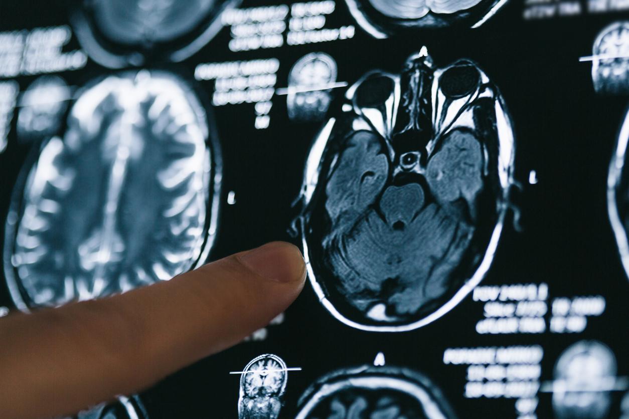
So far, the technique has been tested in patient brains, in vitro, and in mouse models. It involves an examination by two-photon microscope, which is for the moment an obstacle to the development of a simple diagnostic method. But the researchers involved in this discovery are working on an MRI imaging technique to observe the lesions.
.


