Researchers have discovered the origins of “Alice in Wonderland syndrome”.

- The origins of “Alice in Wonderland Syndrome” have been uncovered.
- Concretely, the researchers identified a network encompassing two distinct brain regions: one involved in the perception of the body and the other in the processing of size.
- People with Alice in Wonderland syndrome report episodes in which they perceive parts of their body or objects as too big or too small.
We know more about the origin of the mysterious disease named “Alice in Wonderland Syndrome”.
A new study has in fact just mapped a brain circuit which seems to be involved in this pathology. Concretely, the researchers identified a network encompassing two distinct brain regions: one involved in the perception of the body and the other in the processing of size.
What is Alice in Wonderland syndrome?
In Alice’s Adventures in Wonderland, the heroine follows a white rabbit into its burrow and enters an imaginary world. There she meets a host of emblematic characters and is quickly confronted with a bottle of liquid bearing the inscription “Drink me.” Alice complies and quickly finds herself reduced to a tiny size.
It is this part of the famous novel that gave its name to Alice in Wonderland Syndrome (AIWS). This is a rare phenomenon, of which only 170 cases have so far been described in the scientific literature.
Most often, people with Alice in Wonderland syndrome report episodes in which they perceive parts of their body or objects as too big or too small. In recent years, some researchers have called for broadening the scope of the syndrome to other perceptual disorders such as tachysensitivity (which makes time appear to be passing faster than it should).
With such unusual symptoms and so few documented cases, it has been difficult to determine the cause of AIWS, as the publication’s authors explain. “Alice in Wonderland syndrome has previously been linked to several triggers, such as migraine, brain damage and tumors,” they indicate.
Alice in Wonderland syndrome: specific brain lesions
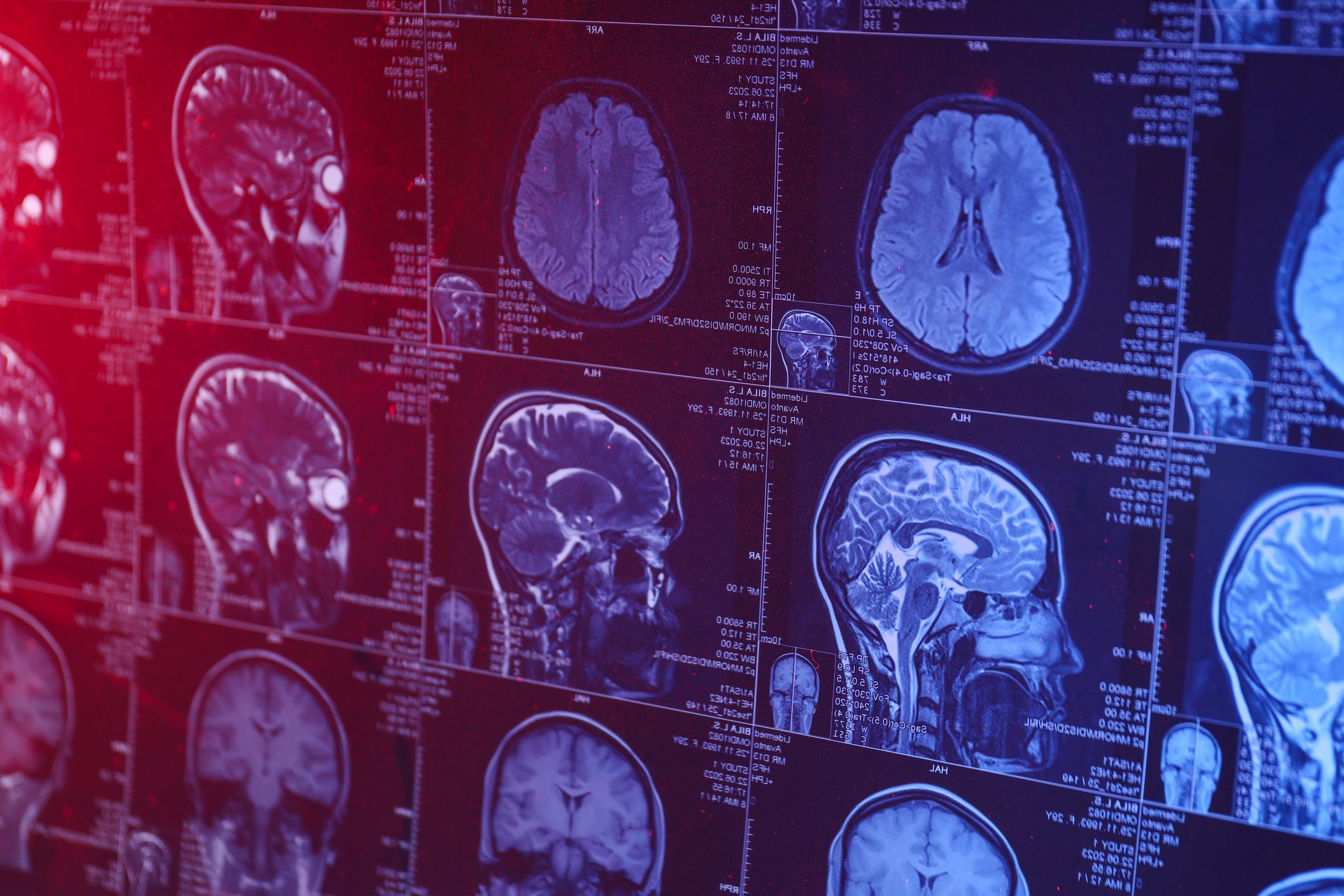
To see more clearly, the team compared the scans of 1,000 healthy people with 1,073 brains with lesions associated with 25 different neuropsychiatric disorders. They then discovered that although the brain lesions present during AIWS varied in terms of location, more than 85% of them were connected to two specific centers: the right extrastriate area (EBA) and the left inferior parietal lobe. (IPL).
The right ABE is part of the visual processing area of the brain and is activated when we observe a body or its parts. As for the left LPI, it is used when we try to determine the size of an object. So it makes sense that these two regions are involved in a disorder that makes body parts appear abnormally small or large.


