The techniques for measuring brain volume would induce a bias, which distorts the understanding of its anatomy and its functioning.

With the advent of neuroscience, the need to study the brain from all angles has greatly arisen. The flagship tool of researchers is the electron microscope. This allows a detailed examination of neuronal cells and brain architecture. Yes but here it is, until today the preparation necessary to study the brain causes it to shrink by at least 30%. The observations made then are somewhat distorted. To remedy this problem, Swiss researchers from the Swiss Federal Institute of Technology in Lausanne (EPFL) have developed a freezing method, described in the newspaper eLife, allowing to keep intact this organ.
For decades, scientists have used aldehyde to “fix” tissue. The purpose of this operation is to stabilize the sample and block all chemical reactions that may take place in the cells. However, this method reduces the size of the brain. The neurons then appear much closer, the structure of the vessels is modified… A problem solved by cryofixation. An innovative process that is not new, since its origin dates back to 1965. This improved method uses jets of liquid nitrogen to instantly freeze brain tissue at -90 ° C. The researchers tested it on samples of the cerebral cortex of mice.
Destroyed connections
Thanks to the 3D electron microscopy photographs, the researchers were able to compare the images of the frozen brain with those of the chemically fixed brain. Analysis showed that the latter is much smaller in volume and exhibits a significant loss of space between neurons. The information would therefore be transmitted very quickly. In addition, it appears that the number of connections between neurons, also called synapses, is lower in the latter than in the cryofigged brain. It is therefore possible that chemical fixation destroys a large number of them. Finally, the researchers also observed that astrocytes, cells surrounding neurons, are much less connected to neuronal cells and blood vessels.

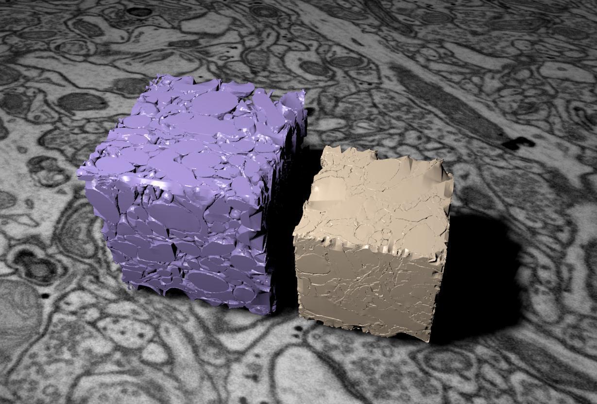
Credit: © Graham Knott / EPFL
To model the compaction of the brain, the researchers used these two cubes. The purple (on the left) represents the actual volume of the brain, preserved by freeze-fixing, and the beige cube (on the right) illustrates the shrinkage caused by the aldehyde.
Preserved anatomy
To assess the impact of these findings, the researchers measured the time it takes for a molecule to travel from one neuron to another in a frozen sample. To their surprise, the times measured in the sample and in functional studies were consistent. An observation which shows that the anatomy of the brain is preserved by cryofixation and deformed by chemical fixation.
“All of this shows us that high pressure freeze-fixation is an ideal method for brain imaging. At the same time, it calls into question previous imaging procedures, which we may need to reexamine in the light of new data, ”explains Graham Knott, co-author of this work. The research team is now focusing on using this technique on other parts of the brain, but also on other types of tissue.
.


