Researchers have identified the stem cells allowing the tapeworm to regenerate. They also understood that the location of the worms close to the head of their host helped them to survive. Eventually, this could lead to new treatments.
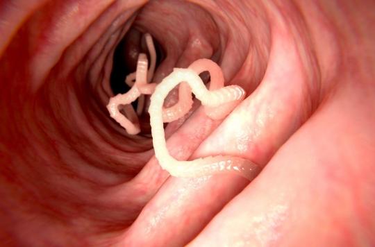
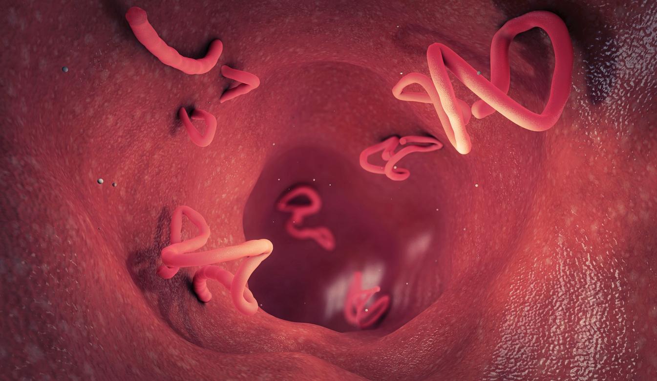
Tapeworms or tapeworms, commonly known as tapeworms, are renowned for the enormous size they can reach and their ability to grow thousands of segments called proglottids. During a normal life cycle, they shed large parts of their body before regenerating to maintain a certain length. However, scientists have never really understood why.
According to a new study published Tuesday, September 24 in the journal eLife, however, American researchers have managed to identify the stem cells that allow them to regenerate. They also understood that the location of the worms close to the head of their host helped them to survive. Ultimately, these findings could explain how tapeworms grow in humans and animals and could lead to new, more effective treatments.
“We know that tapeworm regeneration is likely to involve stem cells, but until now their regenerative potential has never been studied in depth,” said Tania Rozario, from the Morgridge Research Institute of the United States. University of Wisconsin-Madison (USA), which conducted the study. In the latter, “we explored which parts of the tapeworm are able to regenerate and how this regeneration is stimulated by stem cells,” she explains.
Cutting the head of the neck prevents the regeneration of new segments
To answer these questions, Rozario and his colleagues extracted certain fragments of worms which they then bred in the laboratory to determine which regions of the body can regenerate. They thus discovered that neither the head nor the posterior body could regenerate on their own. They were also surprised to find that cutting the head off the neck did not prevent the tapeworm from continuing to grow. On the other hand, the regeneration of new segments was inhibited. Only when the tapeworm’s head and neck were left intact could the tapeworm continuously regenerate into a segment.
The researchers then wanted to see if the neck contained special stem cells that allow tapeworms to regenerate. So they tagged rapidly multiplying cells in the worms and then studied their location in the body. They thus discovered that these cells existed throughout the body and not just the neck.
The researchers therefore deduce that the stem cells are found throughout the tapeworm but that the signals operating only in the neck are necessary to activate them. They therefore had the idea of transferring donor cells from various parts of healthy worms to irradiated, and therefore diseased, flatworms. Result: these cells saved the animals, allowing them to regenerate. In contrast, when the stem cells were removed from the donors, the “rescue” did not take place.
“Location matters a lot”
“It seems that in the case of tapeworms, location matters enormously (…) The head and neck environments provide clues that control the ability of stem cells to regenerate segments, even though the stem cells involved in this process are not confined to one of these regions of the body,” concludes Phillip Newmark, lead author of the study.
When a human catches a tapeworm in their small intestine after eating undercooked meat or raw fish, the first few months after infestation, the growing worm can lead to digestive and appetite disturbances. In the event that the person has consumed pork, it happens that the larval form of the tapeworm leads to cysticercosis. The larvae can then lodge in the muscles, sometimes causing myopathy when the cysts are very numerous, or in the subcutaneous tissues, leading to the formation of painful nodules.
But it is when the larvae localize in the eye or the central nervous system that serious complications can arise. Cerebral cysticercosis, or neurocysticercosis, results in epileptic seizures associated with migraines, nausea and vomiting. The most severely affected people may even develop encephalitis. Other neurological signs are also possible such as meningitis or hydrocephalus (accumulation of cerebrospinal fluid in the spaces of the brain). Regarding ocular cysticercosis, the lesions mainly concern the vitreous and the retina. Untreated, it can progress to blindness.
.


