By performing MRI brain sections, the researchers revealed chronic neuronal inflammation at the periphery of the plaques. Observed in patients with a severe form of multiple sclerosis, this discovery explains the origin of the disease.

- Some patients with a severe form of multiple sclerosis present with lesions with a ring of chronic inflammation around the periphery of the plaques.
- This study, which combines state-of-the-art MRI technique and blood analysis, shows that inflammation, when associated with a high level of neurofilaments in the blood, indicates advanced degeneration of neurons.
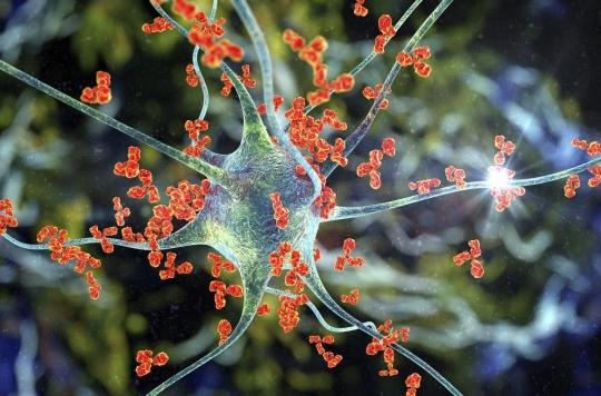
An autoimmune disease of the central nervous system, multiple sclerosis is due to a disruption of the immune system: the latter attacks the brain and nerve fibers by destroying the myelin sheaths responsible for protecting neurons. Gradually, the patients then lose the use of their limbs, have vision, motor and sensitivity disorders.
About ten years ago, research carried out on patients who had developed a severe form of multiple sclerosis revealed a strong chronic inflammation of the plaques. Observed using a high-sensitivity magnetic resonance imaging (MRI) technique, this inflammation results in the appearance of black rings on the periphery of the plaques.
A new international study, led by Dr. Pietro Maggi and involving the universities and university hospitals of Lausanne and Basel (Switzerland), as well as UCLouvain and the Saint-Luc university clinics (Belgium), shows that these black rings are made up of inflammatory cells, including phagocytes, which attack neurons. It has just been published in the journal Neurology.
State-of-the-art technology
The researchers followed 118 patients with multiple sclerosis. Among them, some presented these chronic inflammatory lesions with black rings and others lesions without rings, that is to say which are not yet at the stage of active chronic inflammation. All study participants had their disease status monitored by an MRI scan. They also underwent a blood test to detect the level of neurofilaments, proteins normally present inside neurons.
This technique, which requires the use of an analyzer performing a SIMOA (Single Molecule Array) assay, uses a well plate that can only accommodate a single molecule. Only two machines exist in the French community, including that of the Saint-Luc university clinics, where the work was carried out.
“This technique has been used in neurological diseases to demonstrate neuronal loss, such as in Alzheimer’s degenerative disease, explains Dr. Pietro Maggi, Deputy Head of Clinic of the Neurology Department at Saint-Luc University Clinics. For multiple sclerosis, it has already been used to show that patients with severe forms had a higher level of neurofilaments in the blood, but this is the first time that it has been linked to chronic inflammation. at the cerebral level.
Advanced degeneration of neurons
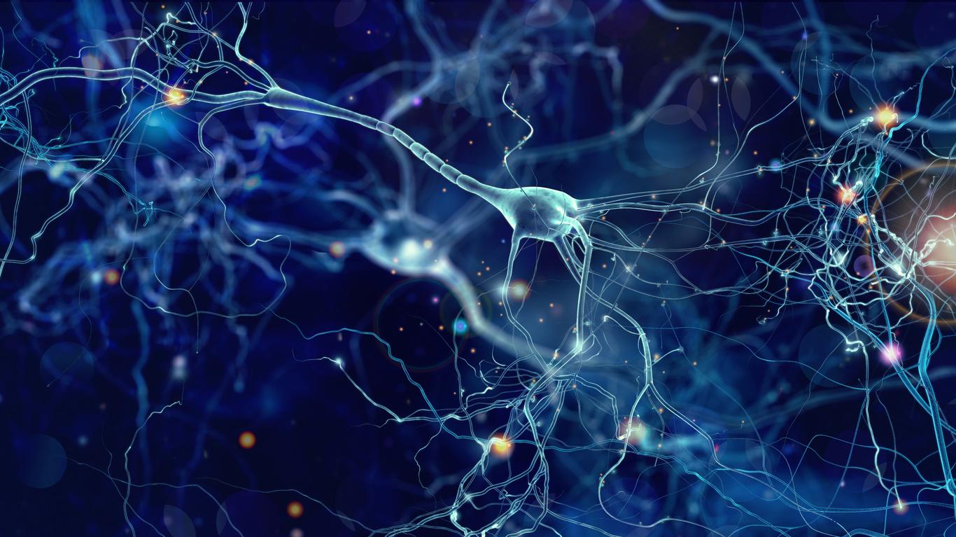
Research has indeed shown a correlation between the presence of lesions with a ring of chronic inflammation on MRI and a significant increase in the level of neurofilaments in the blood, which indicates advanced degeneration of neurons. The presence of lesions with a ring of chronic inflammation on MRI is also associated with more severe clinical disability in patients.
“This is the strongest association ever found at the statistical level, when we look at the variables measured in relation to the increase in neurofilaments in the blood, which means that these chronic active lesions have a primordial role in neurodegeneration. “points out Dr. Maggi.
The results of this study constitute an important advance in the understanding of the pathophysiological mechanisms of multiple sclerosis, believes the researcher. It demonstrates for the first time that the presence of chronic inflammation on MRI is associated with increased neuronal degeneration and a more severe clinical course in patients with multiple sclerosis.
Finally, it shows the possibility of detecting both the presence of chronic cerebral inflammation and its neurodegenerative effect thanks to the combined use of radiological and blood markers.

.


