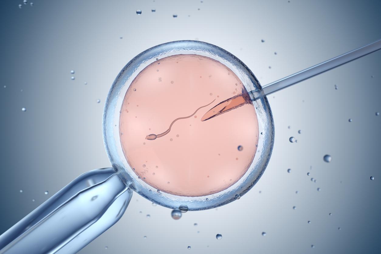
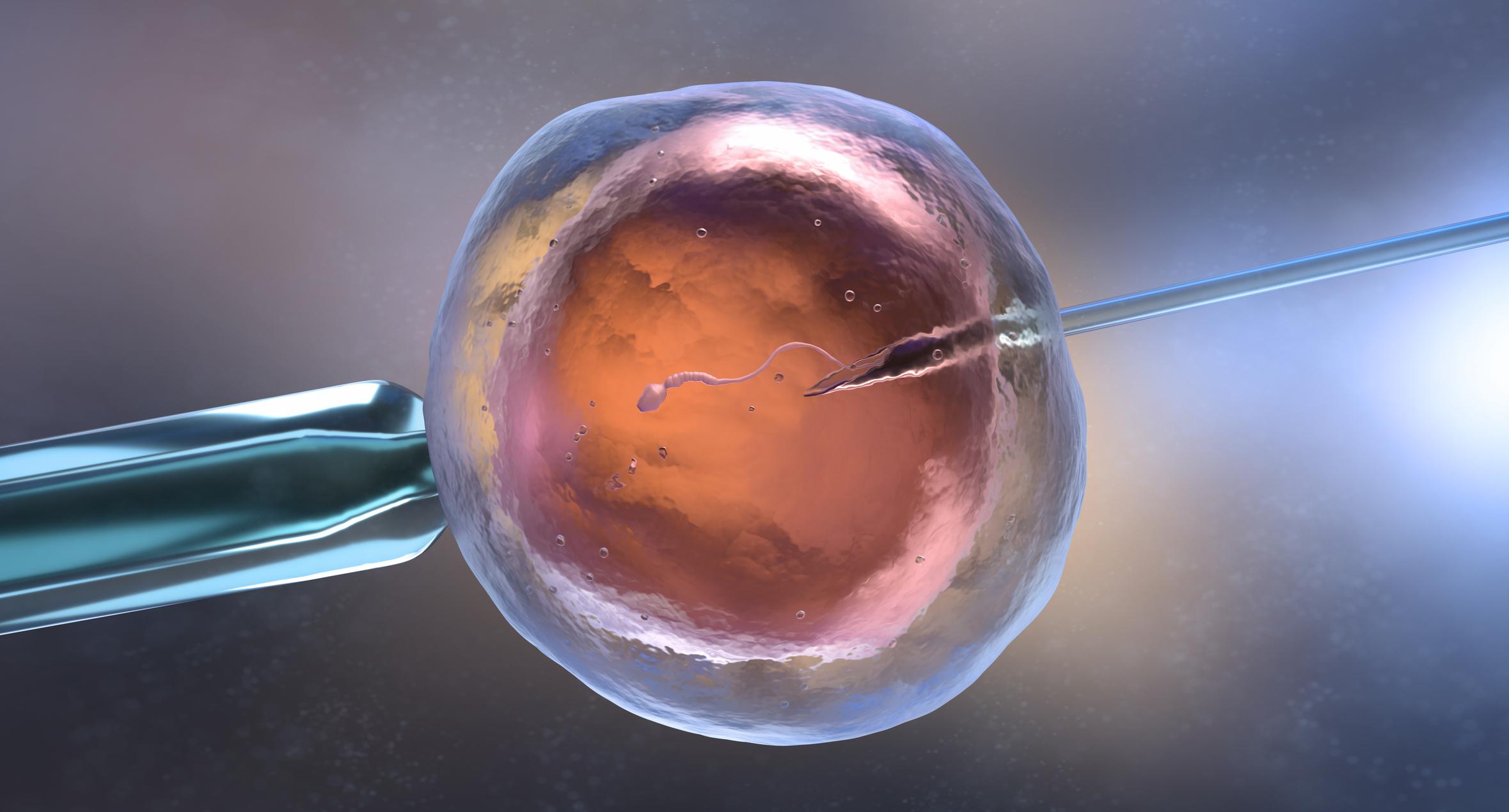
Time-lapse imaging makes it possible to analyze the development of fertilized embryos in vitro before their implantation in the uterus. It could guarantee 73% that the embryos are healthy for a usual rate of 42%.
Researchers from “Care Fertility”, an organization based in the United Kingdom and Ireland, who have developed this technique say ” that they could therefore greatly increase the success rates of pregnancy and live birth, after IVF“.
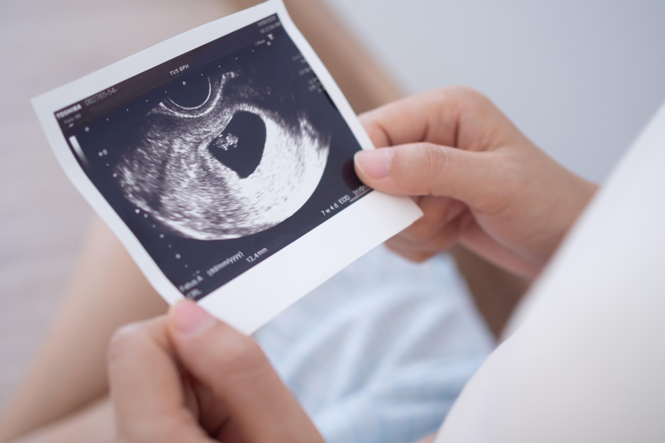
Embryos fertilized in vitro are filmed regularly during their development before being implanted in the uterus. The images provide information necessary to analyze the health and reliability of embryos, in particular the abnormal number of chromosomes (aneuploidy).
To verify the accuracy of this method, the researchers chose 69 couples under IVF.
If usually the embryos are selected after 2 to 6 observations under the microscope, they were, with this method, photographed every 20 minutes.
A more efficient selection
Scientists found that out of 88 embryos, 33 were at low risk for aneuploidy, 51 at medium risk, and 4 at high risk.
Finally, 61% of embryos classified as low risk gave rise to a live birth, 19% of embryos classified as medium risk.
A more efficient selection that increases the success rate. From 42% it goes to 73%. Further studies are necessary to fully validate this new technique.


