An Australian team has recreated a patient’s artery using a supercomputer and 3D printing, in order to facilitate the placement of a stent.
-1456477653.jpg)
Like in the American movie Inner Adventure, being able to visualize perfectly the walls and the structure of the arterial system represents the grail of the cardiologist, in order to deal with myocardial infarctions. Since it is impossible to see precisely what is going on around the heart, researchers at the University of Melbourne came up with the idea of ”taking out” the vessels to examine them. Not directly of course, but by combining an angiogram, a supercomputer and a 3D printer.
“Ideally, we would like to use models to predict which type of stent [petit ressort inséré dans une artère pour la maintenir ouverte, ndlr] we want to ask patients, explains Peter Barlis, cardiologist and professor at the University of Melbourne, responsible for the study. Once our process is running smoothly, we will be able to 3D print a replica of the patient’s artery while they are on the operating table, in order to guide our procedure ”.
To achieve this feat, the researchers perform a simple angiography of the vessels affected by a possible intervention. The images are then processed by a supercomputer which, in less than 24 hours, produces a three-dimensional model. Then, all that remains is to 3D print the arteries, to precisely identify the risk areas with the naked eye, and to choose the appropriate stent.
Treat even before cardiac incident
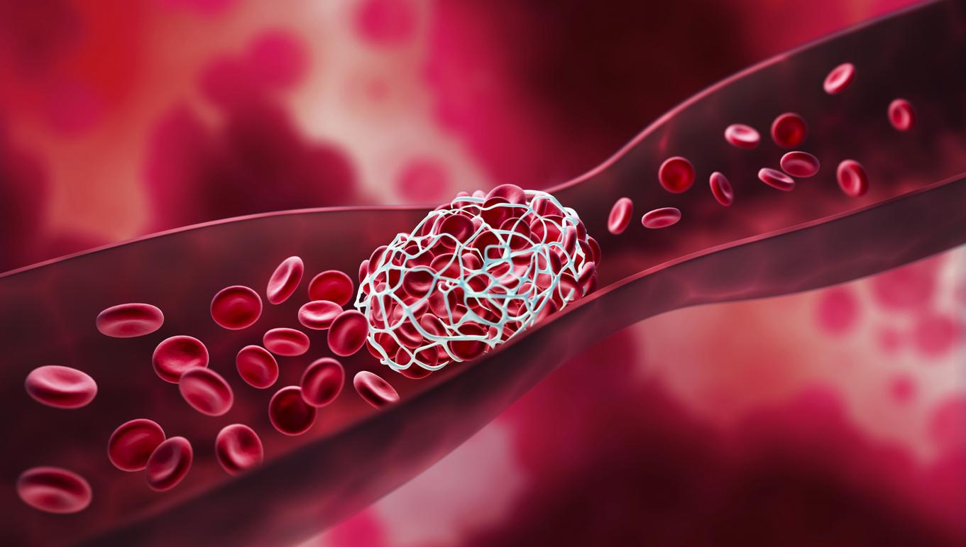
Most serious cardiovascular events are caused by atheromatous plaques, a buildup of lipids, blood products, fatty tissue, and other deposits. When these plaques grow too large or when they break off, they can obstruct a vessel and cause acute coronary syndromes.
At present, it is still difficult to accurately locate these plaques before they become problematic. Even coherence tomographic cameras, which nevertheless have a resolution of less than a micron, are often not sufficient. “If we could identify high-risk plaques more precisely and much earlier, we could take care of infarcts even before they occur,” says Prof. Barlis.
A personalized and absorbable stent
The researchers’ idea lies in the study of blood flow, and in particular of its disturbances. “No two arteries are alike,” explains the cardiologist. A bit like debris that accumulates on the banks of a river, the atheromas cling to specific places. This technique actually gives us a clearer picture of these areas. Everything relies in fact on the computing power of computers which, thanks to the images collected, are able to analyze the interior structure of the vessels and to model it.
Peter Barlis’ team has partnered with the University of Melbourne Engineering School to manufacture viable polymers for 3D printing of stents. Used with the models from the calculator, they could be personalized to perfectly adapt to the morphology of the patients. And to go a little further still, researchers are also interested in special polymers, which would make up absorbable stents capable of delivering drugs directly into the artery.
.


