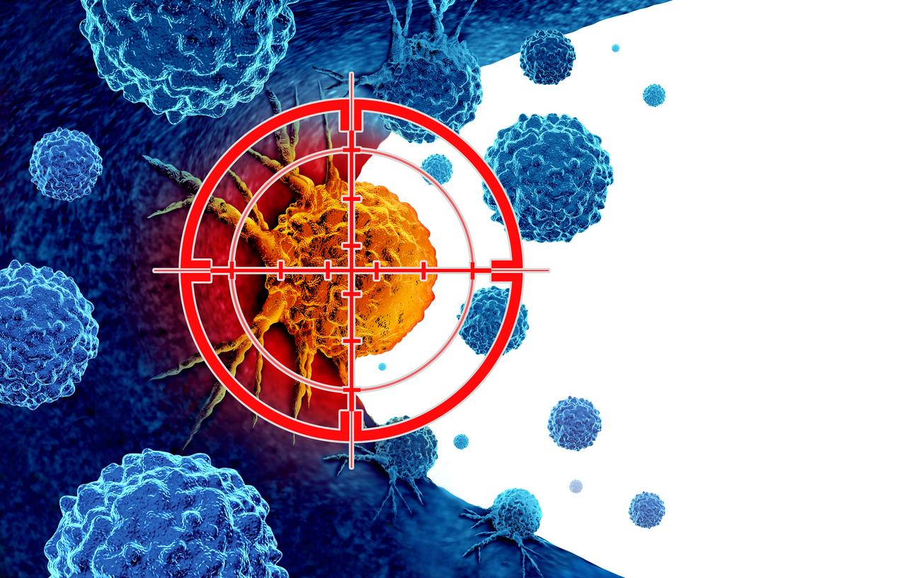
In France, out of the 380,000 cases of cancers diagnosed each year, half are treated by radiotherapy. This technique involves diffusing rays over the tumor area to destroy cancer cells by blocking their ability to multiply. But this process, although effective against cancer, has side effects. Thus, in 5 to 20% of cases, healthy cells close to the tumor are destroyed by radiotherapy: this is radiosensitivity. The National Institute for Health and Medical Research (Inserm) announces the publication of two new studies which provide a better understanding of the response of cells to radiation, and therefore better prevent unwanted effects. These two studies were directed by Nicolas Foray, radiobiologist at Inserm in Lyon.
Focus on the ATM protein
These two studies focused on the ATM protein. When a cell is irradiated, the ATM protein migrates into the cell nucleus and repairs the breaks that radiation has caused in DNA. But if the ATM protein mutates, it is slow to perform this migration. The longer it takes to reach DNA, the higher the radiosensitivity of the cells and the more marked the side effects of radiotherapy. One of the most serious is telangiectasia ataxia, a condition that heavily affects the nervous system and muscles.
ATM migration speed and radiosensitivity are correlated
The first study, published in theInternational Journal of Radiation Biology, is theoretical and looked at the formula that links cell survival and radiation dose. The two authors modeled all the steps in the path of the ATM protein into the cell nucleus. “Thanks to this theoretical study, we can now better understand why a cell is radiosensitive and what the consequences of a failure to recognize or repair double-strand breaks in DNA after irradiation at the cellular level can be.“explains theInserm in a press release.
The second study, published in theInternational Journal of Radiation Oncology, is biological and clinical, and validated the results of the first study. It was carried out on the COPERNIC biological collection, made up of more than a hundred cell lines from radiosensitive patients. The cells were then amplified in the laboratory and irradiated under conditions reproducing those of a radiotherapy session. The researchers then observed how quickly the DNA in these cells was repaired. Then they compared these results to the actual clinical data of each patient from which these cells were taken, in particular the grade of severity of the tissue reactions (ranging from an absence of reaction to the death of the patient, through simple redness and radiation-induced burns. ). Conclusion: “a significant correlation was obtained between the speed of transit of the ATM protein and the grade of severity of the radio-induced tissue reactions“reveals Inserm, and this”regardless of the type of cancer treated or the early or late nature of the adverse tissue reaction after radiotherapy“.
Predictive tests to personalize radiotherapy
This correlation made it possible to define a classification of radiosensitivity in three groups:
– Group I: the ATM protein migrates rapidly = radioresistance
– Group II: delayed ATM migration = moderate radiosensitivity.
– Group III: massive defect in DNA recognition or repair = hyper radiosensitivity
“This classification now makes it possible to anticipate care strategies for radiotherapy treatments.“announces Inserm, specifying that these predictive radiosensitivity tests constitute a promising new tool for better personalizing anti-cancer treatments by radiotherapy, and limiting its undesirable effects.
>> To read also:
Breast cancer: a test to predict the sequelae of radiotherapy
Radiotherapy: antioxidants to protect the skin
Beautiful during and after cancer treatment: essential steps for your skin
The number of breast cancers during pregnancy on the rise


