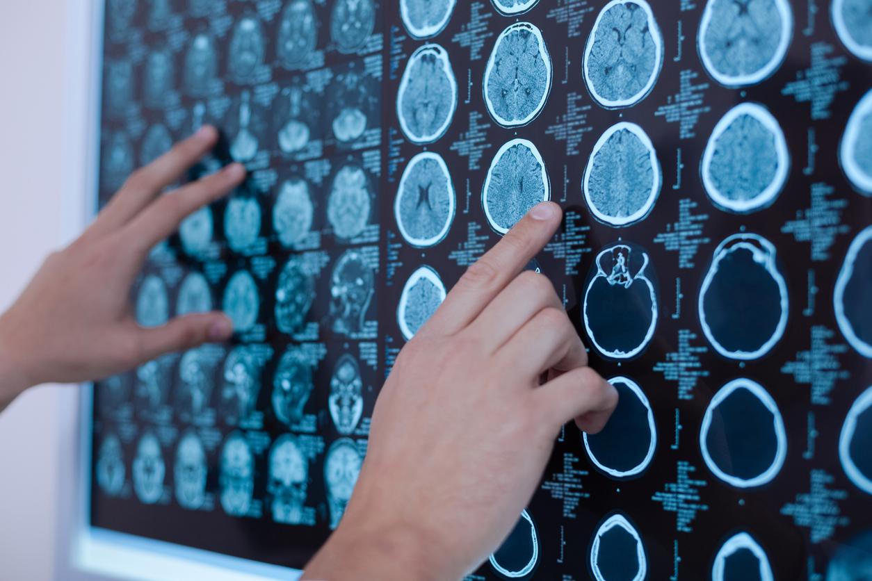
Researchers have developed an algorithm capable of detecting with hitherto unequaled precision and in less than 2.5 minutes the presence of tumors during a patient’s brain operation.

An “accurate and almost real-time preoperative diagnosis of brain tumors”. This is what a new method that combines advanced optical imaging and an artificial intelligence algorithm trained by the analysis of more than 2.5 million biopsy images allows today.
An accurate result in 2 minutes 30
In a study published Monday, January 6 in the journal NatureMedicineresearchers from New York University (NYU) explain that they have succeeded, thanks to this new tool, in establishing an accurate diagnosis at 94.6%, compared to 93.6% when the histological images are analyzed in a conventional manner.
Above all, the algorithm is able to predict in less than 2 minutes 30 whether the cells removed are cancerous or not. What a classic analysis can only reveal between 20 and 30 minutes on average. Thanks to this tool, “we are better equipped to preserve healthy tissue and only remove tissue infiltrated by cancer cells, which translates into fewer complications and better outcomes for cancer patients”, explained to the l ‘OFP neurosurgeon Daniel Orringer. “In neurosurgery and in many other fields of cancer surgery, the detection and diagnosis of tumors during the operation are essential to perform the most appropriate surgical procedure,” he added.
A valuable aid in the detection of brain tumors
Called “stimulated Raman histology” (SRH), this new technique has the ability to reveal tumor infiltration in human tissue by collecting scattered laser light. It thus illuminates essential features that are not typically seen in standard histological images.
To build this new AI tool, the researchers had the algorithm analyze more than 2.5 million tumor samples from 415 patients. These tissue samples were then classified into 13 categories that represent the most common brain tumors. They then recruited 278 patients scheduled to undergo brain tumor resection or epilepsy surgery. Samples of brain tumors from these patients were then biopsied, divided into identical samples and randomly assigned to either the experimental group or the control group.
The microscopic images captured by the imaging were processed and analyzed using the artificial intelligence tool, and in less than two and a half minutes, the surgeons were able to make an accurate diagnosis of brain tumor so far. undetectable, and therefore to be able to remove it.
“The SRH will revolutionize the field of neuropathology by improving decision-making during surgery and providing expert-level assessment in hospitals where trained neuropathologists are unavailable,” asserted in a statement Matija Snuderl, associate professor in the NYU Department of Pathology and co-author of the study.
“NYU Langone’s Department of Neurosurgery has long been a leader in providing the most advanced treatment options to our patients. With (…) this game-changing technology, we are now even better equipped to deliver safe surgeries and quality outcomes for the most complex brain tumor cases,” added Dr. John G. Golfinos, Professor of Neurology and chairman of NYU’s Department of Neurosurgery.

.


