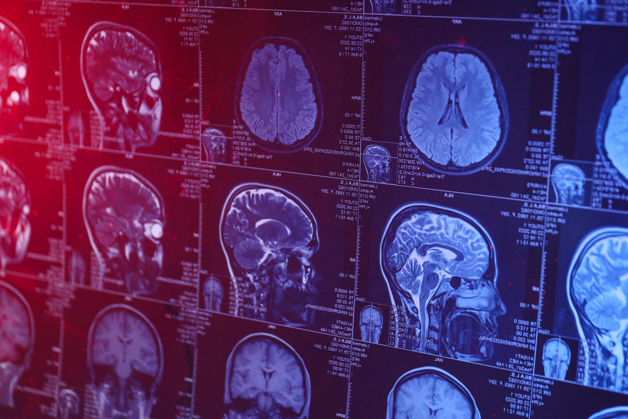
MRI (or Magnetic resonance imaging) can produce so-called quantitative images, i.e. which each map a measurable parameter of the brain (for example blood flow, vascular diameter, etc.). Inserm researchers in collaboration with a research team from the University of Grenoble Alpes have combined various innovative mathematical tools to teach a computer program to analyze these quantitative images from brain MRI and to diagnose possible tumors.
These analyzes showed high reliability results with 100% exact locations and over 90% correct tumor type diagnoses.
How it works ?
First, the program learned to recognize the characteristics of healthy brains. Confronted then with images of brains with cancer, it has thus become able to automatically locate regions whose characteristics diverge from those of healthy tissues and to extract their particularities.
To teach artificial intelligence to differentiate between different types of tumors, the researchers then told it the diagnosis associated with each of the images of diseased brains presented to it.
Read also : Brain cancer: a promising experimental treatment
“This work shows the interest of acquiring this type of images and enlightens radiologists on the analysis tools which they will soon have at their disposal to help them in their interpretations” specifies Emmanuel Barbier, Inserm researcher responsible for the study.
The long-term objective is to succeed in extending the diagnostic potential of this artificial intelligence to other cerebral pathologies, such as Parkinson disease. This innovative method and its results were the subject of a study published in the IEEE-TMI journal.
Read also :
How old is your brain really?
What are the effects of winter on our brains?


