Researchers cut, digitized and 3D reconstructed the brain of a 65-year-old woman. Big brain becomes the ultimate benchmark in terms of anatomy.
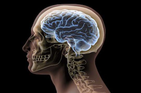
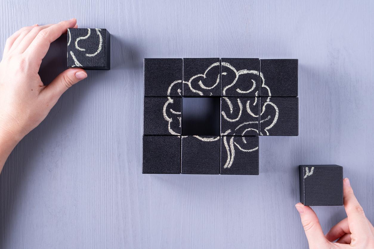
Brain slices thinner than the finest of our hair, this is the feat that a German-Canadian team of researchers has managed to accomplish. Their project, “Big brain”, published on June 21 in the journal Science, is in fact a colossal work. “They cut the brain into thin 20 micron slices and photographed each of those slices. Then they realigned each of these photos to create a large 3D volume, explains Michel Thiebaut de Schotten, researcher at Inserm and author of the “Atlas of human brain connections”.
To date, this level of resolution has only been used to study very small parts of the brain. This is the first time that this technique has been used on an entire brain. The realignment of each of the sections required a very, very large amount of work, ”enthuses the researcher.
How did they “slice the brain”?
The 7404 histological sections actually come from one and the same brain, that of a 65-year-old woman. Each of the cuts was then digitized, a task that alone required around 1,000 hours of work. In fact, “cuts had to be corrected after digitization,” explains Katrin Atmunts, the main author of this work. When cutting the brain, small imperfections occurred on some cuts. It was therefore necessary to control all the images. But these researchers are not their first attempt. They chopped up many brains from corpses before they achieved such a level of finesse. Katrin Amunts and her teams have in fact dedicated the last 15 years to the study of the post-mortem anatomy of the human brain.
What will “Big brain” be used for?
This brain mapping is accessible to everyone. “For the scientific community, this model will become a reference brain allowing a first step towards the unification of scientific research at the microscopic (cellular level) and macroscopic level (large areas activated in functional magnetic resonance imaging). This will be the ultimate reference in terms of anatomy, allowing researchers around the world to make a link between macroscopic observations in living beings thanks to functional imaging and microscopic anatomy of the brain, ”estimates Michel Thiebaut de Schotten. . Moreover, no atlas of this kind had been published for about thirty years. On the other hand, “this will not have any clinical consequence, adds the Inserm researcher because it is a single person brain that cannot quantify normality. “Even if some neuroscientists already imagine that this reconstitution of the brain will help, in the future, to better implant the electrodes for deep brain stimulation interventions. Katrin Atmunts further emphasizes that her work should help interpret the images obtained by MRI.
.


