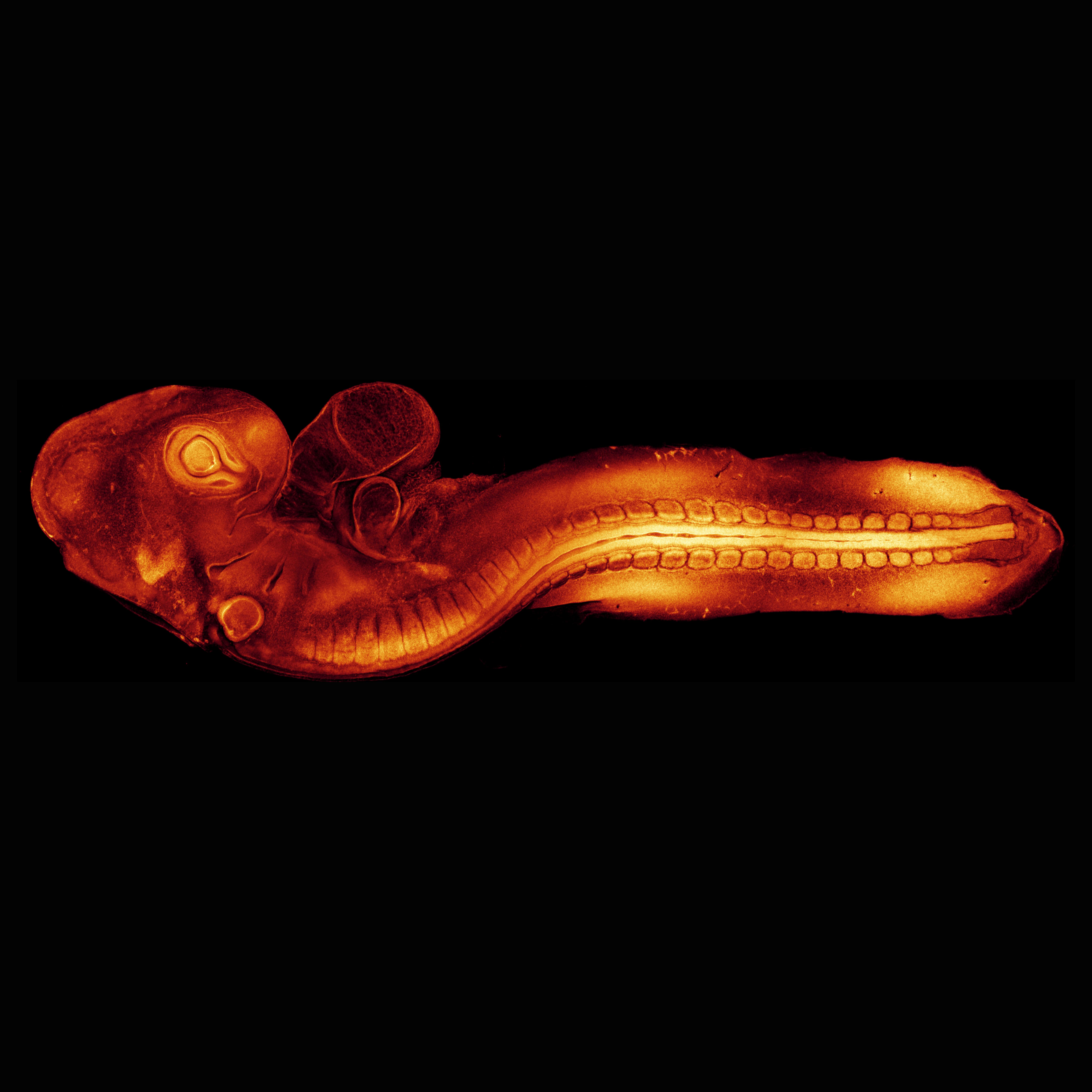
Researchers at the University of Queensland have managed to capture real-time videos of embryonic development.

- Australian researchers have created quails with a fluorescent protein.
- This made it possible to film their fetal development in the egg in real time.
- Researchers have seen how cells move and attach to each other during early embryo development, a discovery that offers insight into birth defects.
Fetal development remains quite mysterious. “Until now, most of our knowledge about post-implantation development has come from studies of static slides, at fixed time points”explains Dr Melanie White from the University of Queensland. The expert and her team have found a way to lift the veil. They have managed to capture for the first time real-time images and videos of early embryonic development. This discovery, published in the journal Journal of Cell Biologycould help understand birth defects.
A fluorescent protein to facilitate the capture of fetal development
To better understand how cells begin to form tissues such as the heart, brain, and spinal cord, scientists used quail eggs. Why this choice? Besides the fact that it is relatively easy to use imaging with an egg, the early development of these birds is very similar to that of a human at the time the embryo implants in the uterus.
The quails in the study had a fluorescent protein that helped reveal the actin cytoskeletons that give cells shape and facilitate their movement. The researchers imaged these eggs and were able to see stages of fetal development never before seen in real time.
“As cells migrate early in development, they produce protrusions called lamellipodia and filopodia, like arms that extend and grip surfaces allowing cells to crawl, or reach around other cells to bring them closer, explains Dr. White. We were able to visualize filopodia from cardiac stem cells deep inside the embryo as they first made contact, sprouting out protrusions and grabbing onto their surroundings and each other to form the early heart.”
“We saw how cells crossed the open neural tube with their protrusions to make contact with the opposite side. The more protrusions the cells formed, the faster the tube closed.”adds the specialist in a press release from his establishment.
Real-time videos to better understand congenital malformations
Seeing fetuses “grow” in real time also allowed the team to better understand the origin of certain congenital malformations. “We saw how cells crossed the open neural tube with their projections to make contact with the opposite side. The more projections the cells formed, the faster the tube closed, explains Dr. White. If this process goes wrong or is disrupted and the tube does not close properly by the fourth week of human development, the embryo will have abnormalities in the brain and spinal cord.”
Having developed this quail model, which makes it easier to study fetal development, the Australian researchers hope to identify proteins or genes that can be targeted or used to screen for congenital malformations.


