


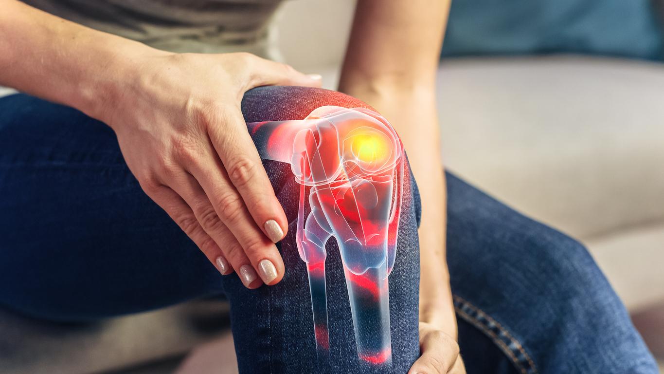
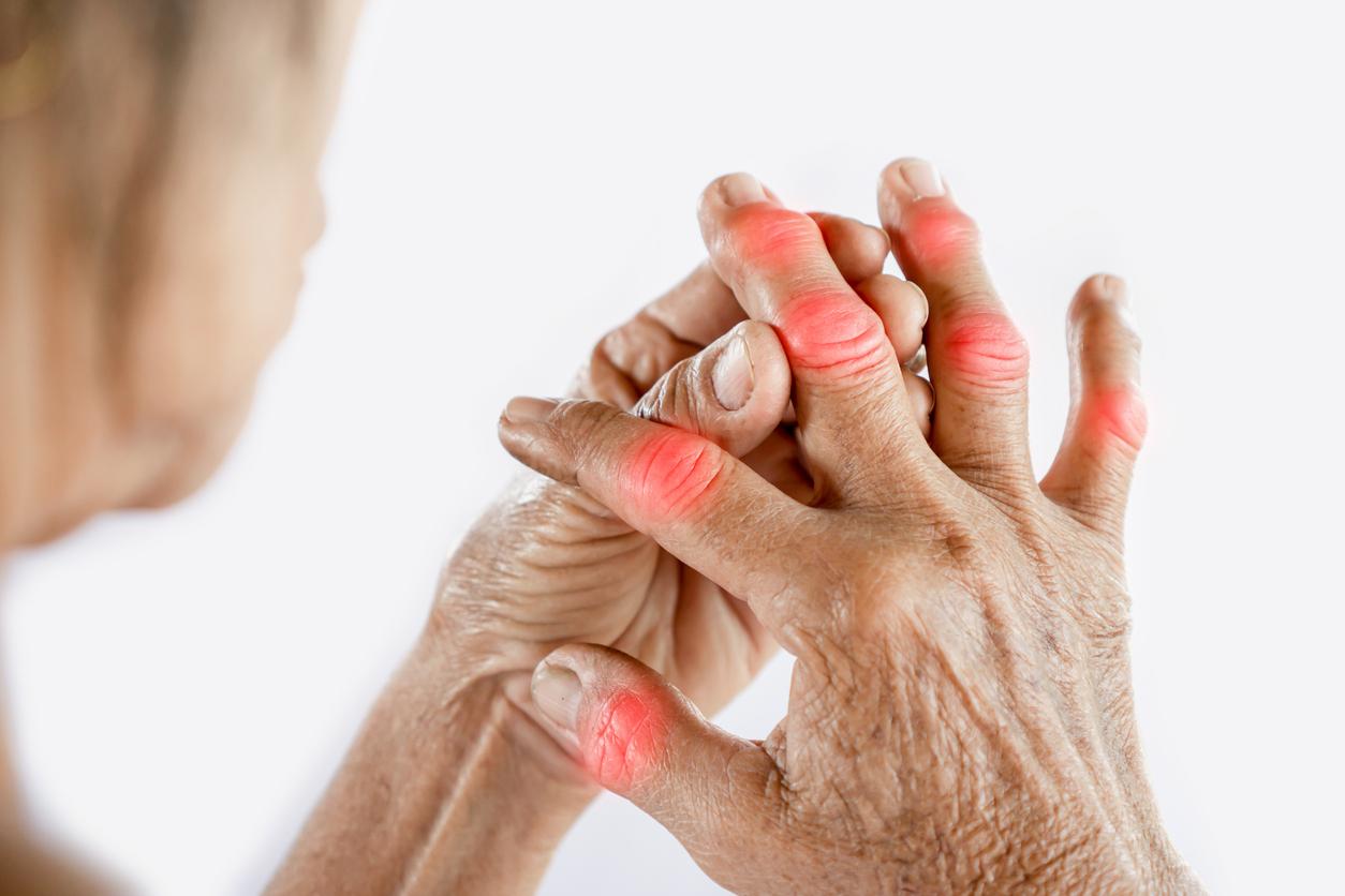
The causes of the appearance of osteoarthritis should be better understood thanks to nanometric medical imaging. It makes it possible to observe the cells of the tissues by which begins the process of degradation of the cartilage which is at the origin of the disease.

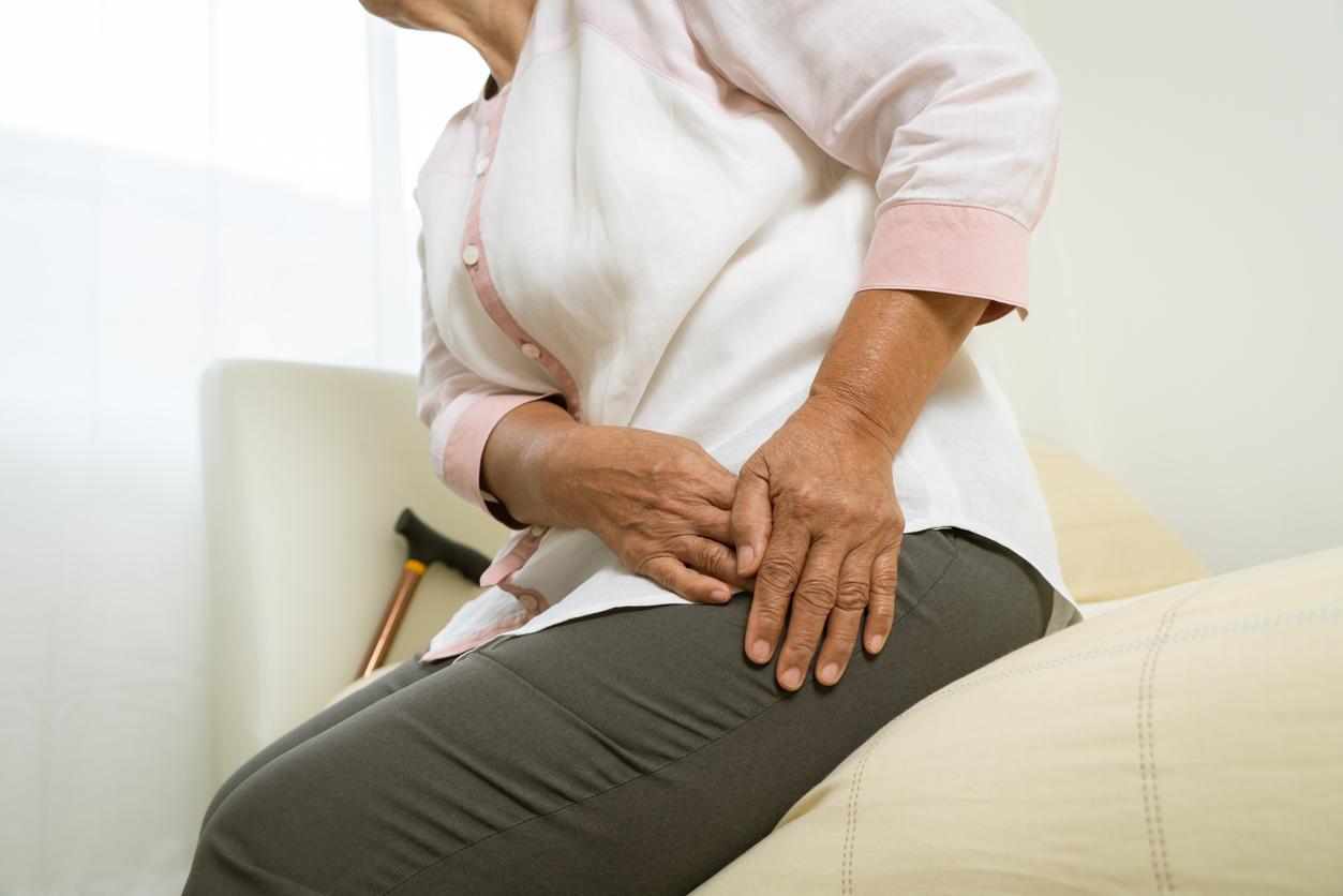
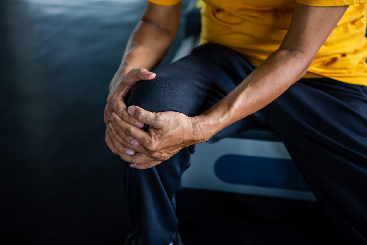
A chronic joint disease, osteoarthritis is most often manifested by mechanical pain and/or discomfort during movement of one or, more rarely, several joints. The risk factors can be aging or mechanical overload of the joint linked to overweight or an architectural anomaly of the joint, but also metabolic abnormalities such as diabetes, or even a hormonal origin or a family predisposition.
“Osteoarthritis will affect most of us throughout our lives, to the point where expensive and difficult surgery to replace the knee or hip joint will often be required after years of disability and pain. ”, said Brian Bay, one of the authors of a new study, published in Biomedical Engineering of Naturewhich opens the door to a better understanding of how the cells that do the work of developing, maintaining and repairing tissues work in a joint.
Cartilage, outer layer of a complex assembly
The cartilage damage that causes osteoarthritis does not become evident until very late in the disease process, because cartilage is simply the outermost layer of a complex array of tissues that lie below the surface. Hence the importance of better observing these tissues where the early changes occur as OA develops, while their basic biomechanical function and the significance of these changes are not yet well understood.
Brian Bay, in collaboration with several scientists, has developed a way to have nanoscale images of complete bones and entire joints. They have developed a sophisticated scanning technique to visualize the “loaded” joints of arthritic and healthy mice, at the ankles, knee or elbow when they are in action, running and walking…
Control cell activity
To do this, they had to improve resolution without compromising field of view, reduce total radiation exposure to preserve tissue mechanics, and prevent motion during scanning. “The use of intact bones and joints means that all functional aspects of the complex layering of tissues are preserved, explains Brian Bay, and the small size of mouse bones leads to imaging that is on the scale cells that grow, maintain and repair tissues.”
“This study for the first time relates measurements of tissue mechanics to the arrangement of the tissues themselves at the cellular level,” said Brian Bay. Interrupting the osteoarthritis process will likely involve controlling cellular activity. This is a breakthrough in linking the clinical problem of joint failure to the most fundamental biological mechanisms involved in maintaining joint health.”
Related work is underway to use these techniques on intervertebral discs and other tissues with high rates of degeneration.

.


